Difference between revisions of "Histology artifacts"
Jump to navigation
Jump to search
| Line 54: | Line 54: | ||
*[[Tissue floater]]. | *[[Tissue floater]]. | ||
*[[Quality]]. | *[[Quality]]. | ||
*[[Foreign material]]. | |||
==References== | ==References== | ||
Revision as of 18:57, 25 November 2014
Histology artifacts are common.
Cautery artifact
Main article: Cautery artifact
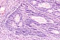
Tissue fold
- Darker well demarcated line with apparent disruption of the architecture.
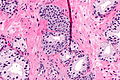
Tissue fold (center of image) in basal cell hyperplasia of the prostate.
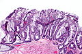
Extensive tissue folding in a SSA.


Chatter artifact
- Fine parallel lines.
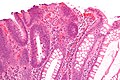
Chatter artifact in tubular adenoma.
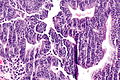
Chatter artifact in complex endometrial hyperplasia.
Chatter artifact in TILs.


Tissue tearing
- Usually due to something intrinsic to the tissue that is hard, e.g. calcium.
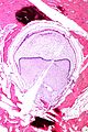
Tearing due to a calcification.

Sponge artifact
- Angulated pieces of tissue with punched-out missing fragments.[1]
- May result in little squares of tissue arranged in a regular pattern.
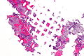
Sponge artifact - very low mag.
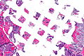
Sponge artifact - low mag.
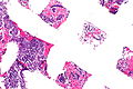
Sponge artifact - intermed. mag.


Bubble artifact
Large
- Large bubbles of air under the cover slip.
Coverslip artifact
Features:
- Black-grey lines - takes on the outline of the tissue beneath it.
- Above the plane-of-focus of the tissue - key feature.
Image:
See also
References
- ↑ Landas SK, Bromley CM (December 1990). "Sponge artifact in biopsy specimens". Arch. Pathol. Lab. Med. 114 (12): 1285–7. PMID 2252429.
- ↑ URL: http://www.statvetlab.com/diagnostic-resources/urinalysis. Accessed on: 3 March 2014.