Difference between revisions of "Histology artifacts"
Jump to navigation
Jump to search
| Line 9: | Line 9: | ||
<gallery> | <gallery> | ||
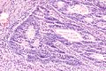
Image:Basal cell hyperplasia of the prostate -- high mag.jpg | Tissue fold (center of image) in [[basal cell hyperplasia of the prostate]]. | Image:Basal cell hyperplasia of the prostate -- high mag.jpg | Tissue fold (center of image) in [[basal cell hyperplasia of the prostate]]. | ||
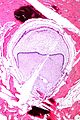
Image: Sessile serrated adenoma -- low mag.jpg | Extensive tissue folding in a [[SSA]]. | |||
</gallery> | </gallery> | ||
Revision as of 01:10, 22 February 2014
Histology artifacts are common.
Cautery artifact
Main article: Cautery artifact
Tissue fold
- Darker well demarcated line with apparent disruption of the architecture.
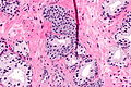
Tissue fold (center of image) in basal cell hyperplasia of the prostate.
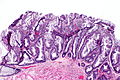
Extensive tissue folding in a SSA.


Chatter artifact
- Fine parallel lines.
Chatter artifact in tubular adenoma.
Chatter artifact in complex endometrial hyperplasia.
Chatter artifact in TILs.


Tissue tearing
- Usually due to something intrinsic to the tissue that is hard, e.g. calcium.
Tearing due to a calcification.

Sponge artifact
- Little squares of tissue in a regular pattern.
Bubble artifact
Large
- Large bubbles of air under the cover slip.