Hibernoma
Jump to navigation
Jump to search
Hibernoma is an uncommon tumour of brown fat. It is an adipocytic tumour.
General
- Consists of brown fat (present in the infants to generate heat).[1]
- Benign.
- Usually asymptomatic.[2]
Epidemiology:
- Young adults.
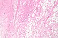
Gross
- Well-circumscribed.
- Lobulated and light-brown on sectioning.
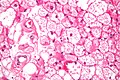
Microscopic
Features:[3]
- Large polygonal/oval cells:
- +/-Prominent blood vessels, central.[6]
DDx:
- Reaction to silicone implant.
Image
Hibernoma - high mag. (WC)
Hibernoma - intermed mag. (WC)
Hibernoma - low mag. (WC)


See also
References
- ↑ Humphrey, Peter A; Dehner, Louis P; Pfeifer, John D (2008). The Washington Manual of Surgical Pathology (1st ed.). Lippincott Williams & Wilkins. pp. 605. ISBN 978-0781765275.
- ↑ Ahmed SA, Schuller I (December 2008). "Pediatric hibernoma: a case review". J. Pediatr. Hematol. Oncol. 30 (12): 900–1. doi:10.1097/MPH.0b013e318184e6dd. PMID 19131775.
- ↑ Chen DY, Wang CM, Chan HL (March 1998). "Hibernoma. Case report and literature review". Dermatol Surg 24 (3): 393–5. PMID 9537018.
- ↑ http://www.pathconsultddx.com/pathCon/diagnosis?pii=S1559-8675(06)70271-6
- ↑ http://surgpathcriteria.stanford.edu/softfat/hibernoma/
- ↑ URL: http://radiographics.rsna.org/content/24/5/1433.full. Accessed on: 11 February 2013.