Difference between revisions of "Hibernoma"
Jump to navigation
Jump to search
(+cat.) |
|||
| (9 intermediate revisions by the same user not shown) | |||
| Line 1: | Line 1: | ||
{{ Infobox diagnosis | |||
| Name = {{PAGENAME}} | |||
| Image = Hibernoma1.jpg | |||
| Width = | |||
| Caption = Hibernoma. [[H&E stain]]. | |||
| Micro = large polygonal/oval cells with central & small nucleus; nucleoli typically prominent; cytoplasm multivacuolated, oval, eosinophilic, granular | |||
| Subtypes = | |||
| LMDDx = reaction to silicone implant | |||
| Stains = | |||
| IHC = | |||
| EM = | |||
| Molecular = | |||
| IF = | |||
| Gross = lobulated lesion, light-brown, usually extremities | |||
| Grossing = | |||
| Site = cervical-supraclavicular, periaortic - both thorax and the abdomen, perirenal; see ''[[soft tissue lesions|soft tissue]]'' - [[adipocytic lesions]] | |||
| Assdx = | |||
| Syndromes = | |||
| Clinicalhx = typically young adults | |||
| Signs = | |||
| Symptoms = | |||
| Prevalence = uncommon | |||
| Bloodwork = | |||
| Rads = | |||
| Endoscopy = | |||
| Prognosis = benign | |||
| Other = | |||
| ClinDDx = [[Lipoma]] | |||
}} | |||
'''Hibernoma''', also '''tumour of [[brown fat]]''',<ref>{{Cite journal | last1 = SHUTE | first1 = D. | title = Tumours of brown fat. | journal = Can Med Assoc J | volume = 71 | issue = 5 | pages = 484-5 | month = Nov | year = 1954 | doi = | PMID = 13209434 }}</ref> is an uncommon [[adipocytic tumour]]. | |||
==General== | |||
*Consists of ''brown fat'' (present in the infants to generate heat).<ref name=Ref_WMSP605>{{Ref WMSP|605}}</ref> | |||
*Benign. | |||
*Usually asymptomatic.<ref name=pmid19131775>{{cite journal |author=Ahmed SA, Schuller I |title=Pediatric hibernoma: a case review |journal=J. Pediatr. Hematol. Oncol. |volume=30 |issue=12 |pages=900–1 |year=2008 |month=December |pmid=19131775 |doi=10.1097/MPH.0b013e318184e6dd |url=}}</ref> | |||
Epidemiology: | |||
*Young adults - disappears with age.<ref name=pmid31281288>{{cite journal |authors=Zoico E, Rubele S, De Caro A, Nori N, Mazzali G, Fantin F, Rossi A, Zamboni M |title=Brown and Beige Adipose Tissue and Aging |journal=Front Endocrinol (Lausanne) |volume=10 |issue= |pages=368 |date=2019 |pmid=31281288 |pmc=6595248 |doi=10.3389/fendo.2019.00368 |url=}}</ref> | |||
==Gross== | |||
*Well-circumscribed. | |||
*Lobulated and light-brown on sectioning. | |||
Locations:<ref name=pmid31281288/> | |||
*Cervical-supraclavicular. | |||
*Periaortic - both thorax and the abdomen. | |||
*Perirenal. | |||
==Microscopic== | |||
Features:<ref name=pmid9537018>{{cite journal |author=Chen DY, Wang CM, Chan HL |title=Hibernoma. Case report and literature review |journal=Dermatol Surg |volume=24 |issue=3 |pages=393–5 |year=1998 |month=March |pmid=9537018 |doi= |url=}}</ref> | |||
*Large polygonal/oval cells: | |||
**Nucleus - central & small.<ref>[http://www.pathconsultddx.com/pathCon/diagnosis?pii=S1559-8675(06)70271-6 http://www.pathconsultddx.com/pathCon/diagnosis?pii=S1559-8675(06)70271-6]</ref> | |||
***Nucleoli typically prominent.<ref>[http://surgpathcriteria.stanford.edu/softfat/hibernoma/ http://surgpathcriteria.stanford.edu/softfat/hibernoma/]</ref> | |||
**Cytoplasm - multivacuolated, oval, eosinophilic, granular. | |||
*+/-Prominent blood vessels, central.<ref>URL: [http://radiographics.rsna.org/content/24/5/1433.full http://radiographics.rsna.org/content/24/5/1433.full]. Accessed on: 11 February 2013.</ref> | |||
DDx: | |||
*Reaction to silicone implant. | |||
===Images=== | |||
<gallery> | |||
Image:Hibernoma1.jpg | Hibernoma - high mag. (WC) | |||
Image:Hibernoma2.jpg | Hibernoma - intermed mag. (WC) | |||
Image:Hibernoma3.jpg | Hibernoma - low mag. (WC) | |||
</gallery> | |||
==See also== | |||
*[[Adipocytic tumours]]. | |||
*[[Lipoma]]. | |||
*[[Brown tumour]]. | |||
==References== | |||
{{Reflist|2}} | |||
[[Category:Adipocytic tumours]] | |||
[[Category:Diagnosis]] | [[Category:Diagnosis]] | ||
Latest revision as of 16:00, 22 April 2024
| Hibernoma | |
|---|---|
| Diagnosis in short | |
 Hibernoma. H&E stain. | |
|
| |
| LM | large polygonal/oval cells with central & small nucleus; nucleoli typically prominent; cytoplasm multivacuolated, oval, eosinophilic, granular |
| LM DDx | reaction to silicone implant |
| Gross | lobulated lesion, light-brown, usually extremities |
| Site | cervical-supraclavicular, periaortic - both thorax and the abdomen, perirenal; see soft tissue - adipocytic lesions |
|
| |
| Clinical history | typically young adults |
| Prevalence | uncommon |
| Prognosis | benign |
| Clin. DDx | Lipoma |
Hibernoma, also tumour of brown fat,[1] is an uncommon adipocytic tumour.
General
- Consists of brown fat (present in the infants to generate heat).[2]
- Benign.
- Usually asymptomatic.[3]
Epidemiology:
- Young adults - disappears with age.[4]
Gross
- Well-circumscribed.
- Lobulated and light-brown on sectioning.
Locations:[4]
- Cervical-supraclavicular.
- Periaortic - both thorax and the abdomen.
- Perirenal.
Microscopic
Features:[5]
- Large polygonal/oval cells:
- +/-Prominent blood vessels, central.[8]
DDx:
- Reaction to silicone implant.
Images
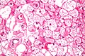
Hibernoma - high mag. (WC)
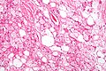
Hibernoma - intermed mag. (WC)
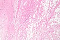
Hibernoma - low mag. (WC)


See also
References
- ↑ SHUTE, D. (Nov 1954). "Tumours of brown fat.". Can Med Assoc J 71 (5): 484-5. PMID 13209434.
- ↑ Humphrey, Peter A; Dehner, Louis P; Pfeifer, John D (2008). The Washington Manual of Surgical Pathology (1st ed.). Lippincott Williams & Wilkins. pp. 605. ISBN 978-0781765275.
- ↑ Ahmed SA, Schuller I (December 2008). "Pediatric hibernoma: a case review". J. Pediatr. Hematol. Oncol. 30 (12): 900–1. doi:10.1097/MPH.0b013e318184e6dd. PMID 19131775.
- ↑ 4.0 4.1 Zoico E, Rubele S, De Caro A, Nori N, Mazzali G, Fantin F, Rossi A, Zamboni M (2019). "Brown and Beige Adipose Tissue and Aging". Front Endocrinol (Lausanne) 10: 368. doi:10.3389/fendo.2019.00368. PMC 6595248. PMID 31281288. https://www.ncbi.nlm.nih.gov/pmc/articles/PMC6595248/.
- ↑ Chen DY, Wang CM, Chan HL (March 1998). "Hibernoma. Case report and literature review". Dermatol Surg 24 (3): 393–5. PMID 9537018.
- ↑ http://www.pathconsultddx.com/pathCon/diagnosis?pii=S1559-8675(06)70271-6
- ↑ http://surgpathcriteria.stanford.edu/softfat/hibernoma/
- ↑ URL: http://radiographics.rsna.org/content/24/5/1433.full. Accessed on: 11 February 2013.