Difference between revisions of "Hemophagocytic syndrome"
Jump to navigation
Jump to search
| Line 11: | Line 11: | ||
| IHC = | | IHC = | ||
| EM = | | EM = | ||
| Molecular = | | Molecular = primary form: FHL1 mutation or FHL2 mutation; autosomal recessive | ||
| IF = | | IF = | ||
| Gross = | | Gross = | ||
| Grossing = | | Grossing = | ||
| Site = [[lymph nodes]], [[liver]], [[spleen]], [[bone marrow]], others | | Site = [[lymph nodes]], [[liver]], [[spleen]], [[bone marrow]], others | ||
| Assdx = only in ''secondary'' (EBV infection, malignancy, rheumatologic disease) | | Assdx = only in ''secondary'' form (EBV infection, malignancy, rheumatologic disease) | ||
| Syndromes = | | Syndromes = | ||
| Clinicalhx = +/-consanguinity for ''primary'' | | Clinicalhx = +/-consanguinity for ''primary'' form | ||
| Signs = fever, [[splenomegaly]], jaundice | | Signs = fever, [[splenomegaly]], jaundice | ||
| Symptoms = | | Symptoms = | ||
Revision as of 05:59, 14 July 2015
| Hemophagocytic syndrome | |
|---|---|
| Diagnosis in short | |
 Micrograph showing phagocytosed RBCs in hemophagocytic syndrome. H&E stain. | |
|
| |
| LM | macrophages containing erythrocytes (red blood cells), leukocytes, platelets |
| Subtypes | primary, secondary |
| LM DDx | overlapping cells, emperipolesis |
| Molecular | primary form: FHL1 mutation or FHL2 mutation; autosomal recessive |
| Site | lymph nodes, liver, spleen, bone marrow, others |
|
| |
| Associated Dx | only in secondary form (EBV infection, malignancy, rheumatologic disease) |
| Clinical history | +/-consanguinity for primary form |
| Signs | fever, splenomegaly, jaundice |
| Prevalence | rare |
| Prognosis | dependent on underlying cause |
Hemophagocytic syndrome, also known as hemophagocytic lymphohistiocytosis, is a rare condition often associated with viral infections.
Clinical
Features:[1]
- Fever.
- Splenomegaly.
- Jaundice.
Involved organs:
Classification
Classified by etiology:[2]
- Primary, i.e. inherited:[3]
- Secondary:
Microscopic
Features:[1]
- Macrophages with phagocytosed:
- Erythrocytes.
- Leukocytes.
- Platelets.
DDx:
- Overlapping cells (red blood cells overlapping macrophages).
- Emperipolesis in the context of other pathology.
Images
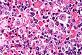
Hemophagocytic syndrome - cropped - very high mag. (WC)
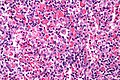
Hemophagocytic syndrome - very high mag. (WC)
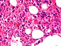
HLH (WC)


www:
See also
References
- ↑ 1.0 1.1 Fisman DN (2000). "Hemophagocytic syndromes and infection". Emerging Infect. Dis. 6 (6): 601–8. PMC 2640913. PMID 11076718. http://www.ncbi.nlm.nih.gov/pmc/articles/PMC2640913/?tool=pubmed.
- ↑ Gupta S, Weitzman S (January 2010). "Primary and secondary hemophagocytic lymphohistiocytosis: clinical features, pathogenesis and therapy". Expert Rev Clin Immunol 6 (1): 137–54. PMID 20383897.
- ↑ Nagai K, Yamamoto K, Fujiwara H, et al. (2010). "Subtypes of familial hemophagocytic lymphohistiocytosis in Japan based on genetic and functional analyses of cytotoxic T lymphocytes". PLoS ONE 5 (11): e14173. doi:10.1371/journal.pone.0014173. PMC 2994802. PMID 21152410. https://www.ncbi.nlm.nih.gov/pmc/articles/PMC2994802/.
- ↑ Online 'Mendelian Inheritance in Man' (OMIM) 603552
- ↑ Online 'Mendelian Inheritance in Man' (OMIM) 603553
- ↑ Humphrey, Peter A; Dehner, Louis P; Pfeifer, John D (2008). The Washington Manual of Surgical Pathology (1st ed.). Lippincott Williams & Wilkins. pp. 576. ISBN 978-0781765275.
- ↑ Jin YK, Xie ZD, Yang S, Lu G, Shen KL (June 2010). "Epstein-Barr virus-associated hemophagocytic lymphohistiocytosis: a retrospective study of 78 pediatric cases in mainland of China". Chin. Med. J. 123 (11): 1426–30. PMID 20819601.