Hemangioma of the liver
Jump to navigation
Jump to search
| Hemangioma of the liver | |
|---|---|
| Diagnosis in short | |
 Cavernous liver hemangioma. H&E stain. | |
| LM DDx | epithelioid hemangioendothelioma, angiosarcoma, metastatic disease |
| Site | liver |
|
| |
| Clinical history | often an incidental finding |
| Symptoms | +/-upper abdominal pain |
| Radiology | well circumscribed mass |
| Prognosis | benign |
| Clin. DDx | metastatic disease |
| Treatment | usually follow-up, non-conservative if very large |
Hemangioma of the liver, also liver hemangioma and hepatic hemangioma, is a benign vascular tumour of the liver, that may be mistaken for metastatic disease.
Hemangiomas, more generally, are dealt with in the hemangioma article.
General
- Benign.[1]
- Usually an incidental finding (incidentaloma) and often asymptomatic.[1]
- Large lesions may present with upper abdominal pain.
Clinical:
- Do not grow in size - can be followed if small or medium size (<10 cm).[1]
Gross
- Variable size.
- Well circumscribed.
Microscopic
Features:
- Channels lined by benign endothelium containing RBCs.
DDx:
Images
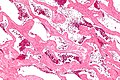
Cavernous liver hemangioma - intermed. mag. (WC/Nephron)
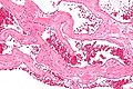
Cavernous liver hemangioma - high mag. (WC/Nephron)

Sign out
Liver Lesion, Core Biopsy:
- Cavernous hemangioma.
- NEGATIVE for malignancy.