Difference between revisions of "Hemangioma of the liver"
Jump to navigation
Jump to search
| Line 69: | Line 69: | ||
- NEGATIVE for malignancy. | - NEGATIVE for malignancy. | ||
</pre> | </pre> | ||
===Micro=== | |||
The sections show dilated vascular spaces containing red blood cells that are lined by endothelial cells without significant atypia. The vascular spaces are separated by bland fibrous tissue. | |||
Abnormal perivascular cells are not identified. The background liver is without atypia and does not have appreciable fat. | |||
==See also== | ==See also== | ||
Revision as of 14:13, 19 October 2015
| Hemangioma of the liver | |
|---|---|
| Diagnosis in short | |
 Cavernous liver hemangioma. H&E stain. | |
| LM DDx | epithelioid hemangioendothelioma, angiosarcoma, metastatic disease |
| Site | liver |
|
| |
| Clinical history | often an incidental finding |
| Symptoms | +/-upper abdominal pain |
| Radiology | well circumscribed mass |
| Prognosis | benign |
| Clin. DDx | metastatic disease |
| Treatment | usually follow-up, non-conservative if very large |
Hemangioma of the liver, also liver hemangioma and hepatic hemangioma, is a benign vascular tumour of the liver, that may be mistaken for metastatic disease.[1]
Hemangiomas, more generally, are dealt with in the hemangioma article.
General
- Benign.[2]
- Can be hard to differentiate from metastatic disease on imaging.[1]
- Usually an incidental finding (incidentaloma) and often asymptomatic.[2]
- Large lesions may present with upper abdominal pain.
- May cause congestive heart failure in infants - if large.[3]
Clinical:
- Do not grow in size - can be followed if small or medium size (<10 cm).[2]
Gross
- Variable size.
- Well circumscribed.
Microscopic
Features:
- Channels lined by benign endothelium containing RBCs.
DDx:
Images
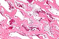
Cavernous liver hemangioma - intermed. mag. (WC/Nephron)
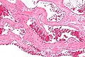
Cavernous liver hemangioma - high mag. (WC/Nephron)

Sign out
Liver Lesion, Core Biopsy:
- Cavernous hemangioma.
- NEGATIVE for malignancy.
Micro
The sections show dilated vascular spaces containing red blood cells that are lined by endothelial cells without significant atypia. The vascular spaces are separated by bland fibrous tissue.
Abnormal perivascular cells are not identified. The background liver is without atypia and does not have appreciable fat.
See also
References
- ↑ 1.0 1.1 Yamashita, Y.; Shimada, M.; Taguchi, K.; Gion, T.; Hasegawa, H.; Utsunomiya, T.; Hamatsu, T.; Matsumata, T. et al. (2000). "Hepatic sclerosing hemangioma mimicking a metastatic liver tumor: report of a case.". Surg Today 30 (9): 849-52. PMID 11039718.
- ↑ 2.0 2.1 2.2 Bajenaru, N.; Balaban, V.; Săvulescu, F.; Campeanu, I.; Patrascu, T. (2015). "Hepatic hemangioma -review.". J Med Life 8 Spec Issue: 4-11. PMID 26361504.
- ↑ Kayaalp, C.; Sabuncuoglu, MZ. (Aug 2015). "Embolization of Liver Hemangiomas.". Hepat Mon 15 (8): e30334. doi:10.5812/hepatmon.30334. PMID 26322113.