Granulomatous prostatitis
Jump to navigation
Jump to search
| Granulomatous prostatitis | |
|---|---|
| Diagnosis in short | |
 Granulomatous prostatitis. H&E stain. | |
|
| |
| LM | granulomas +/- eosinophils |
| Subtypes | non-specific, post-TURP, specific, allergic |
| LM DDx | disseminated granulomatous diseases |
| Stains | GMS stain, Ziehl-Neelsen stain |
| Site | prostate gland |
|
| |
| Prognosis | dependent on underlying etiology |
| Clin. DDx | other types of prostatitis |
Granulomatous prostatitis, also known as prostatic granuloma and prostate gland granuloma, is a common benign finding the prostate.
General
- Common.
- Usually secondary to BCG treatment of bladder cancer.
- Several classifications exist[1] - the most commonly used is by Epstein & Hutchins.
Epstein & Hutchins classification
The groupings:[2]
- Non-specific.
- No cause identified, usu. incidentally discovered.
- Most common.
- Post-TURP.
- Palisading granuloma with necrotic core (histology similar to a rheumatoid nodule[3][4]) +/- eosinophils.
- Specific.
- Identifiable infectious agent, usu. BCG (in the context of treating bladder cancer), rarely tuberculosis and even more rarely various fungi and syphilis.
- Allergic granulomatous prostatitis.
- Usually associated with eosinophils.
- Examples:
Microscopic
Features:
- Granulomas in the prostate - key feature.
- Palisading granulomas with a necrotic core (similar to a rheumatoid nodule) consistent a prior TURP.[3]
- +/-Eosinophils.
Images
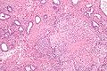
Granulomatous inflammation of the prostate/bladder neck - low mag. (WC/Nephron)
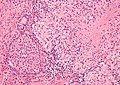
Granulomatous inflammation of the prostate/bladder neck - high mag. (WC/Nephron)

Stains
Note:
- Stains are indicated when there is a suspicion of an infective etiology based on histomorphology or clinical information (e.g. immunosuppression).
Sign out
Post-TURP
PROSTATE GLAND, TRANSURETHRAL RESECTION OF THE PROSTATE (TURP):
- BENIGN PROSTATIC TISSUE WITH GLANDULAR AND STROMAL PROLIFERATION.
- PALISADING GRANULOMA WITH NECROTIC CORE, SEE COMMENT.

COMMENT:
This is morphologically consistent with a post-TURP granuloma.
Idiopathic
A-L. PROSTATE GLAND,
 RIGHT LATERAL SUPERIOR, RIGHT MEDIAL SUPERIOR, RIGHT LATERAL MIDZONE,
 RIGHT MEDIAL MIDZONE, RIGHT LATERAL INTERIOR, RIGHT MEDIAL INFERIOR,
 LEFT LATERAL SUPERIOR, LEFT MEDIAL SUPERIOR, LEFT LATERAL MIDZONE,
 LEFT MEDIAL MIDZONE, LEFT LATERAL INTERIOR, LEFT MEDIAL INFERIOR,
  CORE BIOPSIES:
	- BENIGN PROSTATE TISSUE;
	- GRANULOMATOUS PROSTATITIS, NON-NECROTIZING, SEE COMMENT.

COMMENT:
Granulomatous prostatitis is usually idiopathic. Other possibilities include:
post-procedural granulomatous inflammation (e.g. post-TURP, BCG treatment),
allergic prostatitis and infections.

Infectious etiologies of granulomatous disease should be considered clinically.
See also
References
- ↑ Uzoh, CC.; Uff, JS.; Okeke, AA. (Mar 2007). "Granulomatous prostatitis.". BJU Int 99 (3): 510-2. doi:10.1111/j.1464-410X.2006.06585.x. PMID 17092284.
- ↑ Epstein, JI.; Hutchins, GM. (Sep 1984). "Granulomatous prostatitis: distinction among allergic, nonspecific, and post-transurethral resection lesions.". Hum Pathol 15 (9): 818-25. PMID 6432674.
- ↑ 3.0 3.1 Mies, C.; Balogh, K.; Stadecker, M. (Mar 1984). "Palisading prostate granulomas following surgery.". Am J Surg Pathol 8 (3): 217-21. PMID 6703198.
- ↑ URL: http://www.humpath.com/spip.php?article18010. Accessed on: 26 September 2012.