Granuloma annulare
Jump to navigation
Jump to search
| Granuloma annulare | |
|---|---|
| Diagnosis in short | |
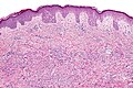
 Granuloma annulare. H&E stain. | |
|
| |
| LM | Palisading granulomas around necrobiotic collagen and mucin |
| Subtypes | superficial (common), deep (not common) |
| LM DDx | rheumatoid nodule, necrobiosis lipoidica, epithelioid sarcoma |
| Stains | alcian blue stain (pH 2.5) |
| Gross | usu. arms and hands, papules |
| Site | skin |
|
| |
| Prognosis | benign |
Granuloma annulare is relatively uncommon problem in dermatopathology.
General
- Benign and self-limited condition.
- Etiology unknown - may be assoc. with trauma.[1]
Gross
- Typically extremities - usu. arms and hands.[1]
Image
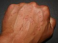
Granuloma annulare. (WC)

Microscopic
Features:[2]
- Dermal palisading granuloma - typically superficial-to-mid dermis - surrounds:
- Necrotic collagen - key feature.
- Nuclei "missing" - have undergone karyolysis.
- Mucin - important.
- Loose/pale, paucicellular, eosinophilic.
- Necrotic collagen - key feature.
- Chronic inflammatory cells.
Notes:
- There may be multiple small foci with intervening normal dermis.[1]
- Granuloma annulare can be subclassified into subcutaneous and interstitial.
- Histomorphologically similar to Rheumatoid nodule.
- Neutrophils may be seen.[3]
DDx:
- Necrobiosis lipoidica - little mucin, no normal dermis between foci,[1] plasma cells - common,[4] may involve the fat - tend to be deeper.
- Rheumatoid nodule - has fibrin in the core of the granuloma (instead of mucin), multinucleated macrophages more common.[5]
- Epithelioid sarcoma - esp. if the lesion appears to be mid-to-deep dermis.
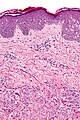
Images
Granuloma annulare - intermed. mag. (WC/Nephron)
Granuloma annulare - high mag. (WC/Nephron)
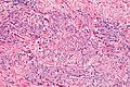
Granuloma annular - palisaded granuloma - high mag. (WC/Nephron)


www:
Stains
- Alcian blue (pH 2.5) +ve (for mucin).[6][1]
Image:
Sign out
SKIN LESION, UPPER BACK, PUNCH BIOPSY:
- SUPERFICIAL PALISADING GRANULOMAS WITH CORES OF NECROBIOTIC COLLAGEN, AND SCANT
  MUCIN; CONSISTENT WITH GRANULOMA ANNULARE.
Skin lesion, left elbow, excision:
- Palisading granulomas with cores of necrobiotic collagen, and scant mucin
  consistent with granuloma annulare.

COMMENT:
An alcian-blue stain (pH 2.5) shows scant mucin.  The granulomas are relatively deep;
however, plasma cells are not apparent. The differential diagnosis is rheumatoid nodule.
Micro
The sections show hair bearing skin with superficial palisading granuloma with cores of necrobiotic collagen and mucin. Plasma cells are not apparent in association with the lesion.
The overlying epidermis matures normally to the surface and has no basal atypia.
See also
References
- ↑ 1.0 1.1 1.2 1.3 1.4 Busam, Klaus J. (2009). Dermatopathology: A Volume in the Foundations in Diagnostic Pathology Series (1st ed.). Saunders. pp. 51. ISBN 978-0443066542.
- ↑ Humphrey, Peter A; Dehner, Louis P; Pfeifer, John D (2008). The Washington Manual of Surgical Pathology (1st ed.). Lippincott Williams & Wilkins. pp. 478. ISBN 978-0781765275.
- ↑ Requena, L.; Fernández-Figueras, MT. (Jun 2007). "Subcutaneous granuloma annulare.". Semin Cutan Med Surg 26 (2): 96-9. doi:10.1016/j.sder.2007.02.006. PMID 17544961.
- ↑ URL: http://dermnetnz.org/pathology/necrobiosis-lipoidica-path.html. Accessed on: 24 January 2012.
- ↑ Busam, Klaus J. (2009). Dermatopathology: A Volume in the Foundations in Diagnostic Pathology Series (1st ed.). Saunders. pp. 52-3. ISBN 978-0443066542.
- ↑ 6.0 6.1 Yun, JH.; Lee, JY.; Kim, MK.; Seo, YJ.; Kim, MH.; Cho, KH.; Kim, MB.; Lee, WS. et al. (May 2009). "Clinical and pathological features of generalized granuloma annulare with their correlation: a retrospective multicenter study in Korea.". Ann Dermatol 21 (2): 113-9. doi:10.5021/ad.2009.21.2.113. PMC 2861218. PMID 20523767. https://www.ncbi.nlm.nih.gov/pmc/articles/PMC2861218/.