Difference between revisions of "Granuloma annulare"
Jump to navigation
Jump to search
(+infobox) |
(→Gross) |
||
| Line 37: | Line 37: | ||
==Gross== | ==Gross== | ||
*Typically extremities - usu. arms and hands.<ref name=Ref_Derm51>{{Ref Derm|51}}</ref> | *Typically extremities - usu. arms and hands.<ref name=Ref_Derm51>{{Ref Derm|51}}</ref> | ||
===Image=== | |||
<gallery> | |||
Image:Granuloma_annulare.jpg | Granuloma annulare. (WC) | |||
</gallery> | |||
==Microscopic== | ==Microscopic== | ||
Revision as of 20:51, 25 June 2013
| Granuloma annulare | |
|---|---|
| Diagnosis in short | |
 Granuloma annulare. H&E stain. | |
|
| |
| LM | Palisading granulomas around necrobiotic collagen and mucin |
| Subtypes | superficial (common), deep (not common) |
| LM DDx | rheumatoid nodule, necrobiosis lipoidica, epithelioid sarcoma |
| Stains | alcian blue stain (pH 2.5) |
| Gross | usu. arms and hands |
| Site | skin |
|
| |
Granuloma annulare is relatively uncommon problem in dermatopathology.
General
- Benign and self-limited condition.
- Etiology unknown - may be assoc. with trauma.[1]
Gross
- Typically extremities - usu. arms and hands.[1]
Image
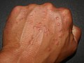
Granuloma annulare. (WC)

Microscopic
Features:[2]
- Dermal palisading granuloma - typically superficial-to-mid dermis - surrounds:
- Necrotic collagen - key feature.
- Nuclei "missing" - have undergone karyolysis.
- Mucin - important.
- Loose/pale, paucicellular, eosinophilic.
- Necrotic collagen - key feature.
- Chronic inflammatory cells.
Notes:
- There may be multiple small foci with intervening normal dermis.[1]
- Granuloma annulare can be subclassified into subcutaneous and interstitial.
- Histomorphologically similar to Rheumatoid nodule.
- Neutrophils may be seen.[3]
DDx:
- Necrobiosis lipoidica - little mucin, no normal dermis between foci,[1] plasma cells - common,[4] may involve the fat - tend to be deeper.
- Rheumatoid nodule - has fibrin in the core of the granuloma (instead of mucin), multinucleated macrophages more common.[5]
- Epithelioid sarcoma - esp. if the lesion appears to be mid-to-deep dermis.
Images
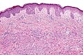
Granuloma annulare - intermed. mag. (WC/Nephron)
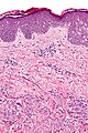
Granuloma annulare - high mag. (WC/Nephron)
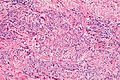
Granuloma annular - palisaded granuloma - high mag. (WC/Nephron)


www:
Stains
- Alcian blue (pH 2.5) +ve (for mucin).[6][1]
Image:
Sign out
Skin lesion, left elbow, excision:
- Palisading granulomas with cores of necrobiotic collagen, and scant mucin
  consistent with granuloma annulare.

COMMENT:
An alcian-blue stain (pH 2.5) shows scant mucin.  The granulomas are relatively deep;
however, plasma cells are not apparent. The differential diagnosis is rheumatoid nodule.
See also
References
- ↑ 1.0 1.1 1.2 1.3 1.4 Busam, Klaus J. (2009). Dermatopathology: A Volume in the Foundations in Diagnostic Pathology Series (1st ed.). Saunders. pp. 51. ISBN 978-0443066542.
- ↑ Humphrey, Peter A; Dehner, Louis P; Pfeifer, John D (2008). The Washington Manual of Surgical Pathology (1st ed.). Lippincott Williams & Wilkins. pp. 478. ISBN 978-0781765275.
- ↑ Requena, L.; Fernández-Figueras, MT. (Jun 2007). "Subcutaneous granuloma annulare.". Semin Cutan Med Surg 26 (2): 96-9. doi:10.1016/j.sder.2007.02.006. PMID 17544961.
- ↑ URL: http://dermnetnz.org/pathology/necrobiosis-lipoidica-path.html. Accessed on: 24 January 2012.
- ↑ Busam, Klaus J. (2009). Dermatopathology: A Volume in the Foundations in Diagnostic Pathology Series (1st ed.). Saunders. pp. 52-3. ISBN 978-0443066542.
- ↑ 6.0 6.1 Yun, JH.; Lee, JY.; Kim, MK.; Seo, YJ.; Kim, MH.; Cho, KH.; Kim, MB.; Lee, WS. et al. (May 2009). "Clinical and pathological features of generalized granuloma annulare with their correlation: a retrospective multicenter study in Korea.". Ann Dermatol 21 (2): 113-9. doi:10.5021/ad.2009.21.2.113. PMC 2861218. PMID 20523767. https://www.ncbi.nlm.nih.gov/pmc/articles/PMC2861218/.