Difference between revisions of "Giant cell tumour of tendon sheath"
Jump to navigation
Jump to search
(redirect) |
(split-out) |
||
| Line 1: | Line 1: | ||
'''Giant cell tumour of tendon sheath''' is a relatively common tumour of small [[joints]]. It is grouped with the [[chondro-osseous tumours]]. It is abbreviated '''GCT of tendon sheath'''. | |||
==General== | |||
*Can be thought of as the small joint version of [[diffuse tenosynovial giant-cell tumour]] ([[AKA]] ''PVNS'').<ref name=Ref_DCHH341>{{Ref DCHH|341}}</ref> | |||
*Rarely recur. | |||
*Classically afflicts the hand.<ref name=Ref_WMSP612>{{Ref WMSP|612}}</ref> | |||
*Rarely malignant.<ref name=pmid19094761>{{Cite journal | last1 = Pan | first1 = YW. | last2 = Huang | first2 = XY. | last3 = You | first3 = JF. | last4 = Tian | first4 = GL. | last5 = Li | first5 = C. | title = [Malignant giant cell tumor of the tendon sheaths in the hand]. | journal = Zhonghua Wai Ke Za Zhi | volume = 46 | issue = 21 | pages = 1645-8 | month = Nov | year = 2008 | doi = | PMID = 19094761 }}</ref><ref name=pmid8230758>{{Cite journal | last1 = Shinjo | first1 = K. | last2 = Miyake | first2 = N. | last3 = Takahashi | first3 = Y. | title = Malignant giant cell tumor of the tendon sheath: an autopsy report and review of the literature. | journal = Jpn J Clin Oncol | volume = 23 | issue = 5 | pages = 317-24 | month = Oct | year = 1993 | doi = | PMID = 8230758 }}</ref> | |||
==Gross== | |||
Features:<ref name=Ref_WMSP612>{{Ref WMSP|612}}</ref> | |||
*Circumscribed mass - yellow-brown to tan. | |||
Note: | |||
*May be associated with bony erosions in larger lesions.<ref name=Ref_WMSP612>{{Ref WMSP|612}}</ref> | |||
Image: | |||
*[http://www.ncbi.nlm.nih.gov/pmc/articles/PMC3122708/figure/Fig2/ GCT of tendon sheath (nih.gov)].<ref name=pmid22282671>{{Cite journal | last1 = Suresh | first1 = SS. | last2 = Zaki | first2 = H. | title = Giant cell tumor of tendon sheath: case series and review of literature. | journal = J Hand Microsurg | volume = 2 | issue = 2 | pages = 67-71 | month = Dec | year = 2010 | doi = 10.1007/s12593-010-0020-9 | PMID = 22282671 }}</ref> | |||
==Microscopic== | |||
Features:<ref name=Ref_DCHH341>{{Ref DCHH|341-2}}</ref> | |||
*Foam cells. | |||
**Cells with moderate to abundant foamy-appearing cytoplasm. | |||
*Multinucleated giant cells - may be scarce. | |||
*+/-Tendon. | |||
**Dense connective tissue. | |||
*+/-Hemosiderin-laden macrophages. | |||
Note: | |||
*Features of malignancy: nuclear pleomorphism,<ref name=pmid8230758>{{Cite journal | last1 = Shinjo | first1 = K. | last2 = Miyake | first2 = N. | last3 = Takahashi | first3 = Y. | title = Malignant giant cell tumor of the tendon sheath: an autopsy report and review of the literature. | journal = Jpn J Clin Oncol | volume = 23 | issue = 5 | pages = 317-24 | month = Oct | year = 1993 | doi = | PMID = 8230758 }}</ref> abnormal mitoses, >10 mitoses/[[HPF]], tumour necrosis lack of maturation to superficial part (nuclei shrink, cytoplasm lipid-ified).<ref name=Ref_DCHH341>{{Ref DCHH|341-2}}</ref> | |||
DDx: | |||
*[[Giant cell lesions]]. | |||
===Images=== | |||
<gallery> | |||
Image:Giant_cell_tumor_of_tendon_sheath_histopathology%281%29.jpg | GCT of tendon sheath. (WC/KGH) | |||
</gallery> | |||
www: | |||
*[http://www.webpathology.com/image.asp?n=1&Case=484 GCT of tendon sheath - very low mag. (webpathology.com)] | |||
*[http://www.webpathology.com/image.asp?case=484&n=5 GCT of tendon sheath - low mag. (webpathology.com)]. | |||
*[http://www.webpathology.com/image.asp?n=4&Case=484 GCT of tendon sheath - high mag. (webpathology.com)]. | |||
==Sign out== | |||
<pre> | |||
LESION, RIGHT INDEX FINGER, EXCISION: | |||
- GIANT CELL TUMOUR OF THE TENDON SHEATH. | |||
</pre> | |||
===Micro=== | |||
The sections show histiocytes and rare multinucleated giant cells on a background of dense connective tissue compatible with tendon. No nuclear atypia is apparent. Rare mitotic activity is identified. No atypical mitoses are apparent. | |||
====Alternate==== | |||
The sections show histiocytes and rare multinucleated giant cells on a background of dense | |||
connective tissue compatible with tendon. Hemosiderin-laden macrophages are present. No | |||
nuclear atypia is apparent. No mitotic activity is apparent. | |||
==See also== | |||
*[[Chondro-osseous tumours]]. | |||
==References== | |||
{{Reflist|2}} | |||
[[Category:Diagnosis]] | [[Category:Diagnosis]] | ||
[[Category:Chondro-osseous tumours]] | |||
Revision as of 22:16, 29 November 2013
Giant cell tumour of tendon sheath is a relatively common tumour of small joints. It is grouped with the chondro-osseous tumours. It is abbreviated GCT of tendon sheath.
General
- Can be thought of as the small joint version of diffuse tenosynovial giant-cell tumour (AKA PVNS).[1]
- Rarely recur.
- Classically afflicts the hand.[2]
- Rarely malignant.[3][4]
Gross
Features:[2]
- Circumscribed mass - yellow-brown to tan.
Note:
- May be associated with bony erosions in larger lesions.[2]
Image:
Microscopic
Features:[1]
- Foam cells.
- Cells with moderate to abundant foamy-appearing cytoplasm.
- Multinucleated giant cells - may be scarce.
- +/-Tendon.
- Dense connective tissue.
- +/-Hemosiderin-laden macrophages.
Note:
- Features of malignancy: nuclear pleomorphism,[4] abnormal mitoses, >10 mitoses/HPF, tumour necrosis lack of maturation to superficial part (nuclei shrink, cytoplasm lipid-ified).[1]
DDx:
Images
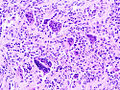
GCT of tendon sheath. (WC/KGH)
.jpg)
www:
- GCT of tendon sheath - very low mag. (webpathology.com)
- GCT of tendon sheath - low mag. (webpathology.com).
- GCT of tendon sheath - high mag. (webpathology.com).
Sign out
LESION, RIGHT INDEX FINGER, EXCISION:
- GIANT CELL TUMOUR OF THE TENDON SHEATH.
Micro
The sections show histiocytes and rare multinucleated giant cells on a background of dense connective tissue compatible with tendon. No nuclear atypia is apparent. Rare mitotic activity is identified. No atypical mitoses are apparent.
Alternate
The sections show histiocytes and rare multinucleated giant cells on a background of dense connective tissue compatible with tendon. Hemosiderin-laden macrophages are present. No nuclear atypia is apparent. No mitotic activity is apparent.
See also
References
- ↑ 1.0 1.1 1.2 Tadrous, Paul.J. Diagnostic Criteria Handbook in Histopathology: A Surgical Pathology Vade Mecum (1st ed.). Wiley. pp. 341. ISBN 978-0470519035.
Cite error: Invalid
<ref>tag; name "Ref_DCHH341" defined multiple times with different content Cite error: Invalid<ref>tag; name "Ref_DCHH341" defined multiple times with different content - ↑ 2.0 2.1 2.2 Humphrey, Peter A; Dehner, Louis P; Pfeifer, John D (2008). The Washington Manual of Surgical Pathology (1st ed.). Lippincott Williams & Wilkins. pp. 612. ISBN 978-0781765275.
- ↑ Pan, YW.; Huang, XY.; You, JF.; Tian, GL.; Li, C. (Nov 2008). "[Malignant giant cell tumor of the tendon sheaths in the hand].". Zhonghua Wai Ke Za Zhi 46 (21): 1645-8. PMID 19094761.
- ↑ 4.0 4.1 Shinjo, K.; Miyake, N.; Takahashi, Y. (Oct 1993). "Malignant giant cell tumor of the tendon sheath: an autopsy report and review of the literature.". Jpn J Clin Oncol 23 (5): 317-24. PMID 8230758.
- ↑ Suresh, SS.; Zaki, H. (Dec 2010). "Giant cell tumor of tendon sheath: case series and review of literature.". J Hand Microsurg 2 (2): 67-71. doi:10.1007/s12593-010-0020-9. PMID 22282671.