Difference between revisions of "Gastric antral vascular ectasia"
Jump to navigation
Jump to search
(fix sp, more images) |
|||
| Line 18: | Line 18: | ||
| Syndromes = | | Syndromes = | ||
| Clinicalhx = 60s or 70s, females:males >2:1 | | Clinicalhx = 60s or 70s, females:males >2:1 | ||
| Signs = | | Signs = +/- melena | ||
| Symptoms = | | Symptoms = | ||
| Prevalence = uncommon | | Prevalence = uncommon | ||
| Bloodwork = usu. [[anemia]] | | Bloodwork = usu. [[anemia]] | ||
| Rads = | | Rads = | ||
| Endoscopy = linear red streaks in antrum - watermelon-like | | Endoscopy = linear red streaks in antrum - watermelon-like | ||
| Line 39: | Line 39: | ||
==General== | ==General== | ||
* | *Etiology not well understood.<ref name=pmid20011731/> | ||
*Typically presents with gastrointestinal bleeding or [[anemia]]. | |||
*Age - usually 60s or 70s. | *Age - usually 60s or 70s. | ||
*Females:males >2:1.<ref name=pmid20011731>{{Cite journal | last1 = Rosenfeld | first1 = G. | last2 = Enns | first2 = R. | title = Argon photocoagulation in the treatment of gastric antral vascular ectasia and radiation proctitis. | journal = Can J Gastroenterol | volume = 23 | issue = 12 | pages = 801-4 | month = Dec | year = 2009 | doi = | PMID = 20011731 | PMC = 2805515 }}</ref> | *Females:males >2:1.<ref name=pmid20011731>{{Cite journal | last1 = Rosenfeld | first1 = G. | last2 = Enns | first2 = R. | title = Argon photocoagulation in the treatment of gastric antral vascular ectasia and radiation proctitis. | journal = Can J Gastroenterol | volume = 23 | issue = 12 | pages = 801-4 | month = Dec | year = 2009 | doi = | PMID = 20011731 | PMC = 2805515 }}</ref> | ||
==Gross/endoscopic appearance== | ==Gross/endoscopic appearance== | ||
* Linear red streaks in antrum - oriented toward the pyloric valve... vaguely resembles a watermelon | * Linear red streaks in antrum - streaks oriented toward the pyloric valve... vaguely resembles a watermelon. | ||
===Endoscopic images=== | |||
<gallery> | <gallery> | ||
Image:Gastric antral vascular ectasia (before and after).png |GAVE. (WC) | Image:Gastric antral vascular ectasia (before and after).png |GAVE. (WC) | ||
</gallery> | </gallery> | ||
www: | |||
*[http://www.pubmedcentral.nih.gov/articlerender.fcgi?artid=2443230&rendertype=figure&id=f1-19 Watermelon stomach (pubmedcentral.nih.gov)]. | |||
==Microscopic== | ==Microscopic== | ||
Features:<ref name=Ref_GLP118>{{Ref GLP|118}}</ref> | Features:<ref name=Ref_GLP118>{{Ref GLP|118}}</ref> | ||
*Fibrin thrombi - '''characteristic feature'''. | *Fibrin thrombi - '''characteristic feature'''. | ||
*Dilated capillaries in lamina propria. | *Dilated capillaries in lamina propria - '''important feature'''. | ||
*+/-Foveollar hyperplasia.<ref name=Ref_GLP119>{{Ref GLP|119}}</ref> | *+/-Foveollar hyperplasia.<ref name=Ref_GLP119>{{Ref GLP|119}}</ref> | ||
Revision as of 17:55, 18 August 2013
| Gastric antral vascular ectasia | |
|---|---|
| Diagnosis in short | |
 Gastric antral vascular ectasia. H&E stain. | |
|
| |
| LM | fibrin thrombi (characteristic feature), dilated capillaries in lamina propria, +/-foveollar hyperplasia |
| LM DDx | portal hypertensive gastropathy |
| Site | stomach |
|
| |
| Clinical history | 60s or 70s, females:males >2:1 |
| Signs | +/- melena |
| Prevalence | uncommon |
| Blood work | usu. anemia |
| Endoscopy | linear red streaks in antrum - watermelon-like |
| Gastric antral vascular ectasia | |
|---|---|
| External resources | |
| Wikipedia | gastric antral vascular ectasia |
Gastric antral vascular ectasia, abbreviated GAVE, is an uncommon pathology of the stomach. It is also known as watermelon stomach due to characteristic endoscopic appearance.[1]
General
- Etiology not well understood.[2]
- Typically presents with gastrointestinal bleeding or anemia.
- Age - usually 60s or 70s.
- Females:males >2:1.[2]
Gross/endoscopic appearance
- Linear red streaks in antrum - streaks oriented toward the pyloric valve... vaguely resembles a watermelon.
Endoscopic images
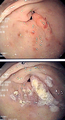
GAVE. (WC)
.png)
www:
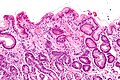
Microscopic
Features:[3]
- Fibrin thrombi - characteristic feature.
- Dilated capillaries in lamina propria - important feature.
- +/-Foveollar hyperplasia.[4]
DDx:
- Portal hypertensive gastropathy - predominantly in the gastric body, usu. associated with cirrhosis, do not have fibrin thrombi.[5]
Images
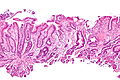
GAVE - intermed. mag. (WC)
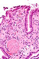
GAVE - very high mag. (WC)


GAVE - low mag. (WC)
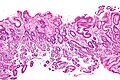
GAVE - intermed. mag. (WC)
GAVE - high mag. (WC)
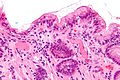
GAVE - two thrombi - very high mag. (WC)


Sign out
STOMACH, BIOPSY:
- GASTRIC ANTRAL VASCULAR ECTASIA WITH FOVEOLAR HYPERPLASIA.
- MILD CHRONIC ACTIVE ANTRAL GASTRITIS.
- NEGATIVE FOR INTESTINAL METAPLASIA.
- NEGATIVE FOR DYSPLASIA.
- NEGATIVE FOR HELICOBACTER ORGANISMS.
Micro
The sections show antral-type gastric mucosa with dilated lamina propria blood vessels and intravascular fibrin thrombi. There is mild foveolar hyperplasia. Numerous neutrophils are present between the foveollar cells and within the lamina propria. Several large clusters of plasma cells are present in the lamina propria.
See also
References
- ↑ Chatterjee S (July 2008). "Watermelon stomach". CMAJ 179 (2): 162. doi:10.1503/cmaj.080461. PMC 2443230. PMID 18625989. http://www.pubmedcentral.nih.gov/articlerender.fcgi?tool=pubmed&pubmedid=18625989.
- ↑ 2.0 2.1 Rosenfeld, G.; Enns, R. (Dec 2009). "Argon photocoagulation in the treatment of gastric antral vascular ectasia and radiation proctitis.". Can J Gastroenterol 23 (12): 801-4. PMC 2805515. PMID 20011731. https://www.ncbi.nlm.nih.gov/pmc/articles/PMC2805515/.
- ↑ Iacobuzio-Donahue, Christine A.; Montgomery, Elizabeth A. (2005). Gastrointestinal and Liver Pathology: A Volume in the Foundations in Diagnostic Pathology Series (1st ed.). Churchill Livingstone. pp. 118. ISBN 978-0443066573.
- ↑ Iacobuzio-Donahue, Christine A.; Montgomery, Elizabeth A. (2005). Gastrointestinal and Liver Pathology: A Volume in the Foundations in Diagnostic Pathology Series (1st ed.). Churchill Livingstone. pp. 119. ISBN 978-0443066573.
- ↑ Iacobuzio-Donahue, Christine A.; Montgomery, Elizabeth A. (2005). Gastrointestinal and Liver Pathology: A Volume in the Foundations in Diagnostic Pathology Series (1st ed.). Churchill Livingstone. pp. 120-1. ISBN 978-0443066573.