Difference between revisions of "Fungi"
(→Microscopic: more) |
|||
| (38 intermediate revisions by 2 users not shown) | |||
| Line 1: | Line 1: | ||
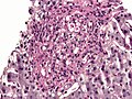
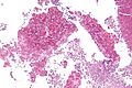
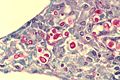
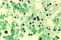
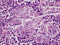
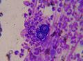
'''Fungi''' are [[microorganisms]] that are occasionally seen by pathologists. | [[Image:Pulmonary aspergillosis.jpg|thumb|right|280px|Fungi (aspergillus). [[H&E stain]].]] | ||
'''Fungi''' (singular '''fungus''') are [[microorganisms]] that are occasionally seen by pathologists. | |||
=Overview= | =Overview= | ||
| Line 15: | Line 16: | ||
*[[Mucor]]. | *[[Mucor]]. | ||
*[[Aspergillus]]. | *[[Aspergillus]]. | ||
===Sign out=== | |||
*The gold standard for determining the microorganisms is culture. | |||
*As anatomical pathologists are approximately 80% accurate (when measured against culture), it is important to state something like ''correlation with culture is recommended''.<ref name=pmid19228642>{{Cite journal | last1 = Sangoi | first1 = AR. | last2 = Rogers | first2 = WM. | last3 = Longacre | first3 = TA. | last4 = Montoya | first4 = JG. | last5 = Baron | first5 = EJ. | last6 = Banaei | first6 = N. | title = Challenges and pitfalls of morphologic identification of fungal infections in histologic and cytologic specimens: a ten-year retrospective review at a single institution. | journal = Am J Clin Pathol | volume = 131 | issue = 3 | pages = 364-75 | month = Mar | year = 2009 | doi = 10.1309/AJCP99OOOZSNISCZ | PMID = 19228642 }}</ref> | |||
==Summary table== | ==Summary table== | ||
{| class="wikitable" | {| class="wikitable sortable" | ||
|- | |- | ||
! Name (disease) | ! Name (disease) | ||
| Line 37: | Line 42: | ||
| ? Immunosuppression | | ? Immunosuppression | ||
| <ref name=Ref_APBR682>{{Ref APBR|682}}</ref> | | <ref name=Ref_APBR682>{{Ref APBR|682}}</ref> | ||
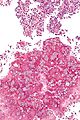
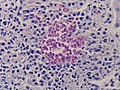
| [ | | [[Image:Pulmonary_aspergillosis.jpg|thumb|center|150px| Aspergillus. (WC)]] | ||
|- | |- | ||
| Zygomycota ([[zygomycosis]]);<br>''more specific''<br>Mucorales (mucormycosis) | | Zygomycota ([[zygomycosis]]);<br>''more specific''<br>Mucorales (mucormycosis) | ||
| Line 47: | Line 52: | ||
| Diabetes, immunodeficient | | Diabetes, immunodeficient | ||
| <ref name=Ref_APBR682>{{Ref APBR|682}}</ref> | | <ref name=Ref_APBR682>{{Ref APBR|682}}</ref> | ||
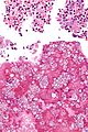
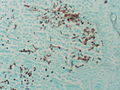
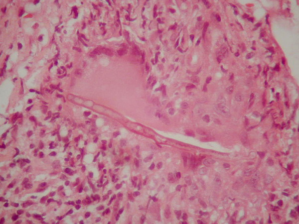
| [ | | [[Image:Zygomycosis.jpg |thumb|center|150px| Zygomycosis. (WC)]] | ||
|- | |- | ||
| Coccidioides, usually C. immitis<br>(coccidioidomycosis) | | Coccidioides, usually C. immitis<br>(coccidioidomycosis) | ||
| Line 57: | Line 62: | ||
| Immunodeficient | | Immunodeficient | ||
| <ref name=Ref_APBR682>{{Ref APBR|682}}</ref> | | <ref name=Ref_APBR682>{{Ref APBR|682}}</ref> | ||
| [http://pathmicro.med.sc.edu/mycology/cocc3.jpg Coccidioidomycosis (med.sc.edu)] [ | | [http://pathmicro.med.sc.edu/mycology/cocc3.jpg Coccidioidomycosis (med.sc.edu)] [[Image:Coccidioides_immitis_on_Sabouraud%27s_medium.jpg |thumb|center|150px|C. immitis (WC)]] | ||
|- | |- | ||
| Histoplasma ([[histoplasmosis]]) | | Histoplasma ([[histoplasmosis]]) | ||
| Line 67: | Line 72: | ||
| Source: soil with bird droppings | | Source: soil with bird droppings | ||
| <ref name=Ref_APBR682>{{Ref APBR|682}}</ref> | | <ref name=Ref_APBR682>{{Ref APBR|682}}</ref> | ||
| [ | | [[Image:Histoplasma_pas-d.jpg|thumb|center|150px| Histoplasmosis. (WC)]] | ||
|- | |- | ||
| Blastomyces ([[blastomycosis]]) | | Blastomyces ([[blastomycosis]]) | ||
| Line 77: | Line 82: | ||
| Habitat: Northeast America, Africa | | Habitat: Northeast America, Africa | ||
| <ref name=Ref_APBR682>{{Ref APBR|682}}</ref><ref>[http://pathmicro.med.sc.edu/mycology/mycology-6.htm http://pathmicro.med.sc.edu/mycology/mycology-6.htm]</ref> | | <ref name=Ref_APBR682>{{Ref APBR|682}}</ref><ref>[http://pathmicro.med.sc.edu/mycology/mycology-6.htm http://pathmicro.med.sc.edu/mycology/mycology-6.htm]</ref> | ||
| [ | | [[Image:Blastomycosis_cropped.JPG | thumb|center|150px|Blastomyces. (WC)]] | ||
|- | |- | ||
| Paracoccidioides ([[paracoccidioidomycosis]]) | | Paracoccidioides ([[paracoccidioidomycosis]]) | ||
| Line 87: | Line 92: | ||
| Clinical??? | | Clinical??? | ||
| <ref name=Ref_APBR682>{{Ref APBR|682}}</ref> | | <ref name=Ref_APBR682>{{Ref APBR|682}}</ref> | ||
| [ | | [[Image:Paracoccidioides_brasiliensis_01.jpg |thumb|center|150px|P. brasiliensis (WC)]] | ||
|- | |- | ||
| Pneumocystis jirovecii ([[pneumocystis carinii pneumonia]]; abbrev. PCP) | | Pneumocystis jirovecii ([[pneumocystis carinii pneumonia]]; abbrev. PCP) | ||
| Line 97: | Line 102: | ||
| [[HIV]]/AIDS associated | | [[HIV]]/AIDS associated | ||
| <ref name=Ref_APBR684>{{Ref APBR|684}}</ref> | | <ref name=Ref_APBR684>{{Ref APBR|684}}</ref> | ||
| [ | | [[Image:Pneumocystosis_carinii_of_lung_in_AIDS_959_lores.jpg|thumb|center|150px| PCP. (WC)]] | ||
|- | |- | ||
| Cryptococcus ([[cryptococcosis]]) | | Cryptococcus ([[cryptococcosis]]) | ||
| Line 107: | Line 112: | ||
| HIV/AIDS associated, most common CNS fungus | | HIV/AIDS associated, most common CNS fungus | ||
| <ref name=Ref_APBR682>{{Ref APBR|682}}</ref> | | <ref name=Ref_APBR682>{{Ref APBR|682}}</ref> | ||
| [ | | [[Image:Cryptococcosis_of_lung_in_patient_with_AIDS._Mucicarmine_stain_962_lores.jpg |thumb|center|150px| Crytococcosis - mucicarmine (WC)]] | ||
|} | |} | ||
Notes: | Notes: | ||
| Line 128: | Line 133: | ||
*** Have a "central dot".<ref name=pathquiz_1>URL: [http://moon.ouhsc.edu/kfung/jty1/opaq/PathQuiz/A6I001-PQ01-M.htm http://moon.ouhsc.edu/kfung/jty1/opaq/PathQuiz/A6I001-PQ01-M.htm]. Accessed on: 19 October 2010</ref> | *** Have a "central dot".<ref name=pathquiz_1>URL: [http://moon.ouhsc.edu/kfung/jty1/opaq/PathQuiz/A6I001-PQ01-M.htm http://moon.ouhsc.edu/kfung/jty1/opaq/PathQuiz/A6I001-PQ01-M.htm]. Accessed on: 19 October 2010</ref> | ||
Images: | ====Images==== | ||
<gallery> | |||
Image:Histoplasma in granuloma pas-d.jpg | Histoplasmosis - granuloma - [[PASD stain]]. (WC) | |||
Image:Histoplasma_pas-d.jpg | Histoplasmosis - [[PASD stain]]. (WC) | |||
Image:Histoplasma_pas-d_small.jpg | Histoplasmosis - cropped - PASD stain. (WC) | |||
Image:Histoplasma_in_granuloma_gms.jpg | Histoplasmosis - [[GMS stain]]. (WC) | |||
</gallery> | |||
www: | |||
*[http://moon.ouhsc.edu/kfung/jty1/opaq/PathQuiz/PQ-Images/A6I001-3.gif The "central dot" of Histoplasma (ouhsc.edu)].<ref name=pathquiz_1/> | *[http://moon.ouhsc.edu/kfung/jty1/opaq/PathQuiz/PQ-Images/A6I001-3.gif The "central dot" of Histoplasma (ouhsc.edu)].<ref name=pathquiz_1/> | ||
*[http://path.upmc.edu/cases/case693.html Histoplasmosis - several images (upmc.edu)]. | *[http://path.upmc.edu/cases/case693.html Histoplasmosis - several images (upmc.edu)]. | ||
| Line 140: | Line 150: | ||
**Usu. from soil. | **Usu. from soil. | ||
*Typical locations: lung, oral cavity.<ref name=Ref_WMSP3>{{Ref WMSP|3}}</ref> | *Typical locations: lung, oral cavity.<ref name=Ref_WMSP3>{{Ref WMSP|3}}</ref> | ||
*+/-Immunodeficiency.<ref name=pmid23824371>{{Cite journal | last1 = Nguyen | first1 = C. | last2 = Barker | first2 = BM. | last3 = Hoover | first3 = S. | last4 = Nix | first4 = DE. | last5 = Ampel | first5 = NM. | last6 = Frelinger | first6 = JA. | last7 = Orbach | first7 = MJ. | last8 = Galgiani | first8 = JN. | title = Recent advances in our understanding of the environmental, epidemiological, immunological, and clinical dimensions of coccidioidomycosis. | journal = Clin Microbiol Rev | volume = 26 | issue = 3 | pages = 505-25 | month = Jul | year = 2013 | doi = 10.1128/CMR.00005-13 | PMID = 23824371 }}</ref> | |||
*Predominantly southwest USA and Mexico.<ref name=pmid23068145>{{Cite journal | last1 = Welsh | first1 = O. | last2 = Vera-Cabrera | first2 = L. | last3 = Rendon | first3 = A. | last4 = Gonzalez | first4 = G. | last5 = Bonifaz | first5 = A. | title = Coccidioidomycosis. | journal = Clin Dermatol | volume = 30 | issue = 6 | pages = 573-91 | month = | year = | doi = 10.1016/j.clindermatol.2012.01.003 | PMID = 23068145 }}</ref> | |||
===Microscopic=== | ===Microscopic=== | ||
| Line 149: | Line 161: | ||
*Spherules may be described as a "bag of marbles". | *Spherules may be described as a "bag of marbles". | ||
Images: | ====Images==== | ||
<gallery> | |||
Image:Coccidioidomycosis - intermed mag.jpg | Coccidioidomycosis - intermed. mag. (WC) | |||
Image:Coccidioidomycosis - high mag.jpg | Coccidioidomycosis - high mag. (WC) | |||
Image:Coccidioidomycosis_-_very_high_mag.jpg | Coccidioidomycosis - very high mag. (WC) | |||
Image:Coccidioidomycosis_-_2_-_gms_-_very_high_mag.jpg | Coccidioidomycosis - GMS - very high mag. (WC) | |||
Image:Mature_spherule_with_endospores_of_Coccidioides_immitis_PHIL_480_lores.jpg | Coccidioides. (WC) | |||
</gallery> | |||
www: | |||
*[http://library.med.utah.edu/WebPath/jpeg2/AIDS092.jpg Coccidioides (med.utah.edu)].<ref>URL: [http://library.med.utah.edu/WebPath/EXAM/IMGQUIZ/pufrm.html http://library.med.utah.edu/WebPath/EXAM/IMGQUIZ/pufrm.html]. Accessed on: 4 December 2011.</ref> | |||
*[http://path.upmc.edu/cases/case516.html Coccidioidomycosis (upmc.edu)]. | |||
==Pneumocystis pneumonia== | ==Pneumocystis pneumonia== | ||
| Line 179: | Line 194: | ||
**Approximately 7-8 μm in size. | **Approximately 7-8 μm in size. | ||
DDx: | |||
*[ | *[[Pulmonary alveolar proteinosis]]. | ||
====Images==== | |||
<gallery> | |||
Image:Pneumocystosis_carinii_of_lung_in_AIDS_959_lores.jpg | The frothy casts of PCP. (WC) | |||
</gallery> | |||
www: | |||
*[http://commons.wikimedia.org/w/index.php?title=Special%3ASearch&search=Pneumocystis+carinii&go=Go Several images of PCP (WC)]. | *[http://commons.wikimedia.org/w/index.php?title=Special%3ASearch&search=Pneumocystis+carinii&go=Go Several images of PCP (WC)]. | ||
| Line 217: | Line 238: | ||
*May be confused with corpora amylacea in the CNS, esp. as they (like cryptococci) stain for methenamine silver, Alcian blue, and PAS.<ref>URL: [http://flylib.com/books/en/2.953.1.17/1/ http://flylib.com/books/en/2.953.1.17/1/]. Accessed on: 15 December 2010.</ref> | *May be confused with corpora amylacea in the CNS, esp. as they (like cryptococci) stain for methenamine silver, Alcian blue, and PAS.<ref>URL: [http://flylib.com/books/en/2.953.1.17/1/ http://flylib.com/books/en/2.953.1.17/1/]. Accessed on: 15 December 2010.</ref> | ||
Images: | ====Images==== | ||
<gallery> | |||
Image:Cryptococcus.jpg | Cryptococcosis - cytology - [[Field stain]]. (WC) | |||
Image:Cryptococcosis_of_lung_in_patient_with_AIDS._Mucicarmine_stain_962_lores.jpg | Crytococcosis - mucicarmine stain. (WC) | |||
Image:Cryptococcosis_of_lung_in_patient_with_AIDS_Methenamine_silver_stain_963_lores.jpg | Crytococcosis - methenamine silver stain. (WC) | |||
</gallery> | |||
www: | |||
*[http://path.upmc.edu/cases/case89/micro.html Cryptococcosis (upmc.edu)]. | |||
*[http://path.upmc.edu/cases/case396.html Cryptococcosis - case 2 (upmc.edu)]. | |||
==Cryptosporidiosis== | ==Cryptosporidiosis== | ||
| Line 236: | Line 259: | ||
**Bluish staining of brush border '''key feature''' - low power. | **Bluish staining of brush border '''key feature''' - low power. | ||
Images | ====Images==== | ||
<gallery> | |||
Image:Cryptosporidiosis_-_very_high_mag.jpg | Cryptosporidiosis - very high mag. (WC) | |||
Image:Cryptosporidiosis_-_very_high_mag_-_cropped.jpg | Cryptosporidosis - very high mag. - cropped (WC) | |||
</gallery> | |||
www: | |||
*[http://www.tulane.edu/~wiser/protozoology/notes/images/cp_path.gif Schematic picture of cryptosporidium & bowel (tulane.edu)]. | |||
*[http://www.brown.edu/Courses/Digital_Path/systemic_path/GI/cryptosporidiosis.html Micrograph of cryptosporidiosis (brown.edu)]. | |||
Notes: | Notes: | ||
| Line 253: | Line 277: | ||
*Yeast forms. | *Yeast forms. | ||
*Locations: oral cavity, vagina. | *Locations: oral cavity, vagina. | ||
===Gross=== | |||
Esophageal candidiasis: | |||
*"Sticky": do not wash-off from the mucosa with water irrigation.<ref>URL: [https://www.ncbi.nlm.nih.gov/books/NBK537268/ https://www.ncbi.nlm.nih.gov/books/NBK537268/]. Accessed on: 2022 June 22.</ref> | |||
===Microscopic=== | ===Microscopic=== | ||
| Line 258: | Line 286: | ||
*''Dimorphic'' - seen in two forms: | *''Dimorphic'' - seen in two forms: | ||
**Pseudohyphae<ref name=Ref_WMSP3>{{Ref WMSP|3}}</ref> - collections of many ''C. albicans'' cells in a branching pattern. | **Pseudohyphae<ref name=Ref_WMSP3>{{Ref WMSP|3}}</ref> - collections of many ''C. albicans'' cells in a branching pattern. | ||
**Yeast form - single cells, 10 to 12 micrometres in diameter | **Yeast form - single cells, 10 to 12 micrometres in diameter.{{fact}} | ||
Notes: | Notes: | ||
*May be described as "sticks and stones". | *May be described as "sticks and stones". | ||
====Images==== | |||
www: | |||
*[http://pathmicro.med.sc.edu/mycology/candi4.jpg Candida (pseudohyphae) - methenamine silver (med.sc.edu)]. | |||
<gallery> | |||
Image:Candida_pap_1.jpg | Candida on Pap test. (WC) | |||
File:Candidiasis (5494228352).jpg | Candidiasis. (WC/Yale Rosen) | |||
File:Candidiasis - PAS (5493634827).jpg | Candidiasis: PAS stain. (WC/Yale Rosen) | |||
File:Candidiasis - GMS (5493634393).jpg | Candidiasis: Pseudohyphae + Budding yeast. (WC/Yale Rosen) | |||
</gallery> | |||
===Stains=== | ===Stains=== | ||
| Line 271: | Line 305: | ||
*PAS +ve. | *PAS +ve. | ||
*Methenamine silver +ve. | *Methenamine silver +ve. | ||
*[[Gram stain|Gram]] +ve.<ref name=pmid20711156>{{Cite journal | last1 = Salerno | first1 = C. | last2 = Pascale | first2 = M. | last3 = Contaldo | first3 = M. | last4 = Esposito | first4 = V. | last5 = Busciolano | first5 = M. | last6 = Milillo | first6 = L. | last7 = Guida | first7 = A. | last8 = Petruzzi | first8 = M. | last9 = Serpico | first9 = R. | title = Candida-associated denture stomatitis. | journal = Med Oral Patol Oral Cir Bucal | volume = 16 | issue = 2 | pages = e139-43 | month = Mar | year = 2011 | doi = | PMID = 20711156 }}</ref> | |||
==Blastomycosis== | ==Blastomycosis== | ||
| Line 285: | Line 320: | ||
*[[Cryptosporidiosis]]. | *[[Cryptosporidiosis]]. | ||
Images | ====Images==== | ||
<gallery> | |||
Image:Blastomycosis_cropped.JPG | Blastomycosis. (WC) | |||
</gallery> | |||
www: | |||
*[http://www.pathguy.com/lectures/blastomycosis.jpg Blastomycosis - budding (pathguy.com)]. | *[http://www.pathguy.com/lectures/blastomycosis.jpg Blastomycosis - budding (pathguy.com)]. | ||
*[http://www.lahey.org/Medical/InfectiousDiseases/ID_Blastomycosis.asp Blastomycosis - with broad budding (lahey.org)]. | *[http://www.lahey.org/Medical/InfectiousDiseases/ID_Blastomycosis.asp Blastomycosis - with broad budding (lahey.org)]. | ||
*[http://pathmicro.med.sc.edu/mycology/blast1.jpg Blastomyces (med.sc.edu)]. | |||
==Mucormycosis== | ==Mucormycosis== | ||
===General=== | ===General=== | ||
| Line 309: | Line 347: | ||
*[[Aspergillosis]]. | *[[Aspergillosis]]. | ||
Images | ====Images==== | ||
<gallery> | |||
Image:Zygomycosis.jpg | Zygomycosis - [[cytology]]. (WC) | |||
</gallery> | |||
www: | |||
*[http://granuloma.homestead.com/ZygoHE_02.jpg Mucormycosis (homestead.com)]. | *[http://granuloma.homestead.com/ZygoHE_02.jpg Mucormycosis (homestead.com)]. | ||
*[http://path.upmc.edu/cases/case181.html Mucormycosis - several images (upmc.edu)]. | *[http://path.upmc.edu/cases/case181.html Mucormycosis - several images (upmc.edu)]. | ||
==Aspergillosis== | ==Aspergillosis== | ||
{{Main|Aspergillosis}} | |||
==Microsporidiosis== | |||
===General=== | ===General=== | ||
* | *A group of (extremely) small intracellular microorganisms - classified as [[fungi]].<ref name=pmid15777637/> | ||
* | **Human pathogenic organisms in this group include: ''Enterocytozoon bieneusi'', ''Encephalitozoon hellem'', and ''Encephalitozoon intestinalis''.<ref name=pmid21844802/> | ||
* | *Important in the context of [[HIV]]/AIDS,<ref name=pmid12775504>{{Cite journal | last1 = Orenstein | first1 = JM. | title = Diagnostic pathology of microsporidiosis. | journal = Ultrastruct Pathol | volume = 27 | issue = 3 | pages = 141-9 | month = | year = | doi = | PMID = 12775504 }}</ref> and solid organ transplant recipients. | ||
*May be seen in immune competent individuals.<ref name=pmid21844802>{{Cite journal | last1 = Didier | first1 = ES. | last2 = Weiss | first2 = LM. | title = Microsporidiosis: not just in AIDS patients. | journal = Curr Opin Infect Dis | volume = 24 | issue = 5 | pages = 490-5 | month = Oct | year = 2011 | doi = 10.1097/QCO.0b013e32834aa152 | PMID = 21844802 }}</ref> | |||
Clinical:<ref name=pmid21844802/> | |||
*Diarrhea. | |||
*Weight loss. | |||
*Abdominal pain. | |||
===Microscopic=== | ===Microscopic=== | ||
Features: | Features: | ||
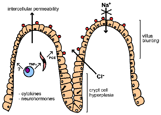
* | *Partial villus atrophy (villous blunting) and crypt hyperplasia.<ref name=pmid21844802/> | ||
* | *Small intracellular microorganisms ~ 1.0-4.0 μm. | ||
Images: | |||
* | *[http://images.wellcome.ac.uk/indexplus/result.html?_IXMAXHITS_=1&_IXACTION_=query&_IXFIRST_=5&_IXemailreal=true&_IXbox=50007&_IXSPFX_=templates%2Ft&_IXFPFX_=templates%2Ft Microsporidiosis (wellcome.ac.uk)]. | ||
*[http://wwwnc.cdc.gov/eid/article/18/2/11-1319-f1.htm Microsporidiosis (cdc.gov)].<ref>URL: [http://wwwnc.cdc.gov/eid/article/18/2/11-1319_article.htm http://wwwnc.cdc.gov/eid/article/18/2/11-1319_article.htm]. Accessed on: 2 June 2012.</ref> | |||
===EM=== | |||
*Small intracellular microorganisms ~ 1.0-4.0 μm.<ref name=pmid15777637>{{Cite journal | last1 = Didier | first1 = ES. | title = Microsporidiosis: an emerging and opportunistic infection in humans and animals. | journal = Acta Trop | volume = 94 | issue = 1 | pages = 61-76 | month = Apr | year = 2005 | doi = 10.1016/j.actatropica.2005.01.010 | PMID = 15777637 }}</ref> | |||
* | |||
Image: | |||
* | *[http://commons.wikimedia.org/wiki/File:Fibrillanosema_spore.jpg Microsporidium (WC)].{{fact}} | ||
=See also= | =See also= | ||
Latest revision as of 15:03, 22 June 2022

Fungi (singular fungus) are microorganisms that are occasionally seen by pathologists.
Overview
- There are lots of 'em. Below are a few of 'em.
Terminology:[1]
- Hyphae = microscopic filamentous growth (of fungi) -- single cell.
- Mycelial = filamentous network of hyphae.
- Septae/septation = hyphae may be subdivided by septae -- if they aren't they are one mass of protoplasm. (?)
- Dimorphism = exist in two forms; e.g. single cell (yeast) and mycelial growth.
- Pseudohyphae = looks like hyphae --but branching pattern is created by separate cells.[2]
Tissue invasive fungi
Typically:[3]
Sign out
- The gold standard for determining the microorganisms is culture.
- As anatomical pathologists are approximately 80% accurate (when measured against culture), it is important to state something like correlation with culture is recommended.[4]
Summary table
| Name (disease) | Kingdom | Size | Shape | Stains | Other (microscopic) | Clinical | References | Image |
|---|---|---|---|---|---|---|---|---|
| Aspergillus (aspergillosis) | Fungi | ? | Hyphae that branching with 45 degrees angle |
PAS-D | Fruiting heads when aerobic | ? Immunosuppression | [5] | |
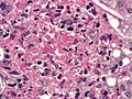
| Zygomycota (zygomycosis); more specific Mucorales (mucormycosis) |
Fungi | ? | Branching hyphae with variable width | ? | Granulomata assoc. | Diabetes, immunodeficient | [5] | |
| Coccidioides, usually C. immitis (coccidioidomycosis) |
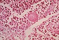
Fungi | Large - 20-60 micrometers, endospores 1-5 micrometers |
Spherules | Stains? | Other? | Immunodeficient | [5] | Coccidioidomycosis (med.sc.edu) |
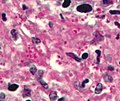
| Histoplasma (histoplasmosis) | Fungi | 2-5 micrometers | Spherical | GMS | Intracellular (unlike candida), granulomata | Source: soil with bird droppings | [5] | |
| Blastomyces (blastomycosis) | Fungi | 5-15 micrometres | Spherical (yeast) | Stains? | Granulomas, broad-based budding yeast | Habitat: Northeast America, Africa | [5][6] | |
| Paracoccidioides (paracoccidioidomycosis) | Fungi | 6-60 micrometres | Spherical (yeast) | Stains? | Multiple budding "steering wheel" appearance | Clinical??? | [5] | |
| Pneumocystis jirovecii (pneumocystis carinii pneumonia; abbrev. PCP) | Fungi (previously thought to be a protozoan) | 7-8 micrometres | "Dented ping-pong ball" | GMS | Usually in clusters of alveolar casts with a honeycomb appearance | HIV/AIDS associated | [7] | |
| Cryptococcus (cryptococcosis) | Fungi | 5-15 micrometres | Yeast | GMS | Prominent (i.e. thick polysaccharide) capsule | HIV/AIDS associated, most common CNS fungus | [5] |


Notes:
- Bold text = key features.
Specific fungi
Histoplasmosis
General
- Organism: Histoplasma.
- Specific organism: Histoplasma capulatum.
- Typical location: lung.
- Common in immunosuppressed individuals, e.g. HIV/AIDS population.
- Extrapulmonary or disseminated histoplasmosis is considered to be AIDS-defining.[8]
Microscopic
Features:
- Often in yeast form - in tissue, spherical, 2-5 micrometres.[9]
- Intracellular[10] - may be within macrophages that form a granuloma.
- Nice bright red on PAS-D.
- Have a "central dot".[11]
- Nice bright red on PAS-D.
Images
Histoplasmosis - granuloma - PASD stain. (WC)
Histoplasmosis - PASD stain. (WC)
Histoplasmosis - cropped - PASD stain. (WC)
Histoplasmosis - GMS stain. (WC)


www:
Coccidioidomycosis
General
- Organism: Coccidioides.
- Specific organism: Coccidioides immitis.
- Usu. from soil.
- Typical locations: lung, oral cavity.[12]
- +/-Immunodeficiency.[13]
- Predominantly southwest USA and Mexico.[14]
Microscopic
Features:
- Forms spherules 60-80 μm in size.[9]
- Contain endospores 1-5 μm in diameter.
Notes:
- Spherules may be described as a "bag of marbles".
Images
Coccidioidomycosis - intermed. mag. (WC)
Coccidioidomycosis - high mag. (WC)
Coccidioidomycosis - very high mag. (WC)
Coccidioidomycosis - GMS - very high mag. (WC)
Coccidioides. (WC)


www:
Pneumocystis pneumonia
- Abbreviated PCP.
- AKA Pneumocystis jirovecii pneumonia.
General
- Organism: pneumocystis,
- Specific organism: Pneumocystis jirovecii[16] (used to be called Pneumocystis carinii).
- May be spelled Pneumocystis jiroveci.
- Fungus... used to be considered a parasite.
- Typical location: lung.
Clinical:
- Opportunistic infection - typically in HIV +ve individuals.
- May have subtle findings on chest X-ray.
Microscopic
Features:
- Form frothy aggregates that take the shape of the alveoli they sit within, i.e. they form "alveolar casts".
- "Dented ping-pong ball" appearance.[9] **Remember PCP = ping-pong.
- Approximately 7-8 μm in size.
DDx:
Images
The frothy casts of PCP. (WC)
www:
Stains
- GMS stain +ve.
Cryptococcosis
General
- Organism: Cryptococcus.
- Specific organism: C. neoformans.
- Opportunistic infection.
- Typical location: lung.
- Most common fungus seen in CSF specimens.[5]
Trivia:
- Crypto- = hidden/invisible.[17]
- Why the name? A. The capsule is almost invisible.
Gross
Features (brain):
- Small cystic spaces, often diffuse.
- Known as "soap bubble brain".
Image:
Microscopic
Features:
- Yeast:
- Usually accompanied by very little inflammation.[18]
Notes:
- May be confused with corpora amylacea in the CNS, esp. as they (like cryptococci) stain for methenamine silver, Alcian blue, and PAS.[19]
Images
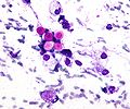
Cryptococcosis - cytology - Field stain. (WC)
Crytococcosis - mucicarmine stain. (WC)
Crytococcosis - methenamine silver stain. (WC)


www:
Cryptosporidiosis
General
- Caused by cryptosporidium.
- Fecal-oral transmission.
- Usu. in immunoincompetent individuals, e.g. HIV/AIDS.
Microscopic
Features:
- Uniform spherical nodules 2-4 micrometres in diameter, typical location - GI tract brush border.
- Bluish staining of brush border key feature - low power.
Images
Cryptosporidiosis - very high mag. (WC)
Cryptosporidosis - very high mag. - cropped (WC)


www:
- Schematic picture of cryptosporidium & bowel (tulane.edu).
- Micrograph of cryptosporidiosis (brown.edu).
Notes:
- Cryptosporidium parvum?[20]
Candidiasis
- In the context of pap tests see: Gynecologic_cytopathology#Candida.
General
- Commonly Candida albicans.
- Yeast forms.
- Locations: oral cavity, vagina.
Gross
Esophageal candidiasis:
- "Sticky": do not wash-off from the mucosa with water irrigation.[21]
Microscopic
Features:
- Dimorphic - seen in two forms:
- Pseudohyphae[12] - collections of many C. albicans cells in a branching pattern.
- Yeast form - single cells, 10 to 12 micrometres in diameter.[citation needed]
Notes:
- May be described as "sticks and stones".
Images
www:
Candida on Pap test. (WC)
Candidiasis. (WC/Yale Rosen)
Candidiasis: PAS stain. (WC/Yale Rosen)
Candidiasis: Pseudohyphae + Budding yeast. (WC/Yale Rosen)

.jpg)
.jpg)
.jpg)
Stains
Features:
Blastomycosis
General
- Usually Blastomyces dermatitidis - fungus.
- May be in the oral cavity.[12]
Microscopic
Features:
- Broad-based budding yeast -- is Blastomyces.[23]
- The interface between two separating fungi, i.e. fungi in the process of reproducing, is very large.
DDx:
Images
Blastomycosis. (WC)
www:
- Blastomycosis - budding (pathguy.com).
- Blastomycosis - with broad budding (lahey.org).
- Blastomyces (med.sc.edu).
Mucormycosis
General
- Causative organism: Mucorales.
- Kingdom: Fungi.
- AKA Zygomycota (zygomycosis).
- Associated with diabetes, immunodeficiency.
Microscopic
Features:[5]
- Branching hyphae with variable width.
- Granulomata associated.
Notes:
- Not septated.
- Branching angle typically ~90 degrees.
DDx:
Images
Zygomycosis - cytology. (WC)
www:
Aspergillosis
Microsporidiosis
General
- A group of (extremely) small intracellular microorganisms - classified as fungi.[24]
- Human pathogenic organisms in this group include: Enterocytozoon bieneusi, Encephalitozoon hellem, and Encephalitozoon intestinalis.[25]
- Important in the context of HIV/AIDS,[26] and solid organ transplant recipients.
- May be seen in immune competent individuals.[25]
Clinical:[25]
- Diarrhea.
- Weight loss.
- Abdominal pain.
Microscopic
Features:
- Partial villus atrophy (villous blunting) and crypt hyperplasia.[25]
- Small intracellular microorganisms ~ 1.0-4.0 μm.
Images:
EM
- Small intracellular microorganisms ~ 1.0-4.0 μm.[24]
Image:
See also
References
- ↑ http://www.fungionline.org.uk/1intro/3growth_forms.html
- ↑ http://pathmicro.med.sc.edu/mycology/mycology-3.htm
- ↑ CM 17 Apr 2009.
- ↑ Sangoi, AR.; Rogers, WM.; Longacre, TA.; Montoya, JG.; Baron, EJ.; Banaei, N. (Mar 2009). "Challenges and pitfalls of morphologic identification of fungal infections in histologic and cytologic specimens: a ten-year retrospective review at a single institution.". Am J Clin Pathol 131 (3): 364-75. doi:10.1309/AJCP99OOOZSNISCZ. PMID 19228642.
- ↑ 5.00 5.01 5.02 5.03 5.04 5.05 5.06 5.07 5.08 5.09 5.10 Lefkowitch, Jay H. (2006). Anatomic Pathology Board Review (1st ed.). Saunders. pp. 682. ISBN 978-1416025887.
- ↑ http://pathmicro.med.sc.edu/mycology/mycology-6.htm
- ↑ Lefkowitch, Jay H. (2006). Anatomic Pathology Board Review (1st ed.). Saunders. pp. 684. ISBN 978-1416025887.
- ↑ Schneider E, Whitmore S, Glynn KM, Dominguez K, Mitsch A, McKenna MT (December 2008). "Revised surveillance case definitions for HIV infection among adults, adolescents, and children aged <18 months and for HIV infection and AIDS among children aged 18 months to <13 years--United States, 2008". MMWR Recomm Rep 57 (RR-10): 1–12. PMID 19052530. http://www.cdc.gov/mmwr/preview/mmwrhtml/rr5710a1.htm.
- ↑ 9.0 9.1 9.2 Humphrey, Peter A; Dehner, Louis P; Pfeifer, John D (2008). The Washington Manual of Surgical Pathology (1st ed.). Lippincott Williams & Wilkins. pp. 103. ISBN 978-0781765275.
- ↑ Gorocica, P.; Taylor, ML.; Alvarado-Vásquez, N.; Pérez-Torres, A.; Lascurain, R.; Zenteno, E. (May 2009). "The interaction between Histoplasma capsulatum cell wall carbohydrates and host components: relevance in the immunomodulatory role of histoplasmosis.". Mem Inst Oswaldo Cruz 104 (3): 492-6. PMID 19547878.
- ↑ 11.0 11.1 URL: http://moon.ouhsc.edu/kfung/jty1/opaq/PathQuiz/A6I001-PQ01-M.htm. Accessed on: 19 October 2010
- ↑ 12.0 12.1 12.2 Humphrey, Peter A; Dehner, Louis P; Pfeifer, John D (2008). The Washington Manual of Surgical Pathology (1st ed.). Lippincott Williams & Wilkins. pp. 3. ISBN 978-0781765275.
- ↑ Nguyen, C.; Barker, BM.; Hoover, S.; Nix, DE.; Ampel, NM.; Frelinger, JA.; Orbach, MJ.; Galgiani, JN. (Jul 2013). "Recent advances in our understanding of the environmental, epidemiological, immunological, and clinical dimensions of coccidioidomycosis.". Clin Microbiol Rev 26 (3): 505-25. doi:10.1128/CMR.00005-13. PMID 23824371.
- ↑ Welsh, O.; Vera-Cabrera, L.; Rendon, A.; Gonzalez, G.; Bonifaz, A.. "Coccidioidomycosis.". Clin Dermatol 30 (6): 573-91. doi:10.1016/j.clindermatol.2012.01.003. PMID 23068145.
- ↑ URL: http://library.med.utah.edu/WebPath/EXAM/IMGQUIZ/pufrm.html. Accessed on: 4 December 2011.
- ↑ Redhead, SA.; Cushion, MT.; Frenkel, JK.; Stringer, JR.. "Pneumocystis and Trypanosoma cruzi: nomenclature and typifications.". J Eukaryot Microbiol 53 (1): 2-11. doi:10.1111/j.1550-7408.2005.00072.x. PMID 16441572.
- ↑ URL: http://en.wiktionary.org/wiki/crypto-. Accessed on: 12 April 2012.
- ↑ Lefkowitch, Jay H. (2006). Anatomic Pathology Board Review (1st ed.). Saunders. pp. 423 Q29. ISBN 978-1416025887.
- ↑ URL: http://flylib.com/books/en/2.953.1.17/1/. Accessed on: 15 December 2010.
- ↑ http://www.dpd.cdc.gov/dpdx/HTML/Cryptosporidiosis.htm
- ↑ URL: https://www.ncbi.nlm.nih.gov/books/NBK537268/. Accessed on: 2022 June 22.
- ↑ Salerno, C.; Pascale, M.; Contaldo, M.; Esposito, V.; Busciolano, M.; Milillo, L.; Guida, A.; Petruzzi, M. et al. (Mar 2011). "Candida-associated denture stomatitis.". Med Oral Patol Oral Cir Bucal 16 (2): e139-43. PMID 20711156.
- ↑ Veligandla, SR.; Hinrichs, SH.; Rupp, ME.; Lien, EA.; Neff, JR.; Iwen, PC. (Oct 2002). "Delayed diagnosis of osseous blastomycosis in two patients following environmental exposure in nonendemic areas.". Am J Clin Pathol 118 (4): 536-41. doi:10.1309/JEJ0-3N98-C3G8-21DE. PMID 12375640.
- ↑ 24.0 24.1 Didier, ES. (Apr 2005). "Microsporidiosis: an emerging and opportunistic infection in humans and animals.". Acta Trop 94 (1): 61-76. doi:10.1016/j.actatropica.2005.01.010. PMID 15777637.
- ↑ 25.0 25.1 25.2 25.3 Didier, ES.; Weiss, LM. (Oct 2011). "Microsporidiosis: not just in AIDS patients.". Curr Opin Infect Dis 24 (5): 490-5. doi:10.1097/QCO.0b013e32834aa152. PMID 21844802.
- ↑ Orenstein, JM.. "Diagnostic pathology of microsporidiosis.". Ultrastruct Pathol 27 (3): 141-9. PMID 12775504.
- ↑ URL: http://wwwnc.cdc.gov/eid/article/18/2/11-1319_article.htm. Accessed on: 2 June 2012.