Difference between revisions of "Fulminant hepatic necrosis"
(create) |
(split out) |
||
| Line 1: | Line 1: | ||
'''Fulminant hepatic necrosis''' is extensive cell death in the liver. It is classified as a [[medical liver disease]]. | |||
===General=== | |||
Etiology: | |||
*Viral, i.e. [[Hepatitis A]], [[Hepatitis B]]; [[Hepatitis C]] - extremely rare. | |||
*Trauma. | |||
*Shock. | |||
===Microscopic=== | |||
Features: | |||
*Hepatocyte [[necrosis]]. | |||
*Bile duct proliferation. | |||
DDx: | |||
*[[Cholangiocarcinoma]]. | |||
*[[Angiosarcoma]]. | |||
===Images=== | |||
A. [[File:1 Necrosis 2 680x512px.tif| Submassive hepatic necrosis.]] | |||
<br> | |||
B. [[File:2 Necrosis 2 680x512px.tif| Submassive hepatic necrosis.]] | |||
<br> | |||
C. [[File:3 Necrosis 2 680x512px.tif| Submassive hepatic necrosis.]] | |||
<br> | |||
D. [[File:4 Necrosis 2 680x512px.tif| Submassive hepatic necrosis.]] | |||
<br> | |||
E. [[File:5 Necrosis 2 680x512px.tif| Submassive hepatic necrosis.]] | |||
<br> | |||
F. [[File:6 Necrosis 2 680x512px.tif| Submassive hepatic necrosis.]] | |||
<br> | |||
Submassive hepatic necrosis. Patient had transaminases in the thousands that rapidly dropped to normal. A/ Pink preserved parenchyma strews empty necrotic spaces. B. Focus of necrosis with no apparent hepatocytes macrophages abuts apparently normal liver. C. Iron stain shows the macrophages bear hemosiderin. D. Reticulin stain highlights the recently dead liver cells. E. Reticulin stain shows a necrotic bridge forming; the multiple small black circles preclude diagnosis of a fibrous bridge. F. Trichrome shows the necrotic bridge (“collapse”) lacks much collagen deposition, as would be expected for bridging fibrosis. | |||
==See also== | |||
*[[Necrosis]]. | |||
*[[Medical liver disease]]. | |||
==References== | |||
{{Reflist|1}} | |||
[[Category:Diagnosis]] | [[Category:Diagnosis]] | ||
[[Category:Medical liver disease]] | |||
Revision as of 14:46, 18 June 2017
Fulminant hepatic necrosis is extensive cell death in the liver. It is classified as a medical liver disease.
General
Etiology:
- Viral, i.e. Hepatitis A, Hepatitis B; Hepatitis C - extremely rare.
- Trauma.
- Shock.
Microscopic
Features:
- Hepatocyte necrosis.
- Bile duct proliferation.
DDx:
Images
A. 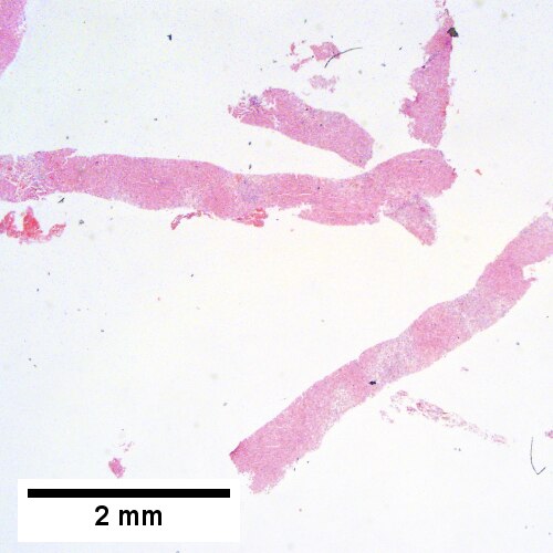
B. 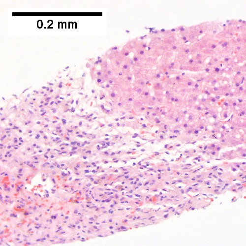
C. 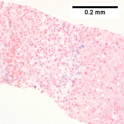
D. 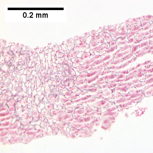
E. 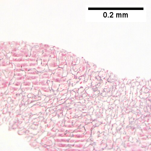
F. 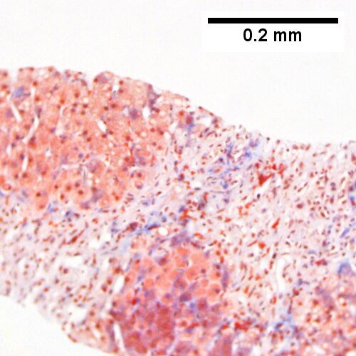
Submassive hepatic necrosis. Patient had transaminases in the thousands that rapidly dropped to normal. A/ Pink preserved parenchyma strews empty necrotic spaces. B. Focus of necrosis with no apparent hepatocytes macrophages abuts apparently normal liver. C. Iron stain shows the macrophages bear hemosiderin. D. Reticulin stain highlights the recently dead liver cells. E. Reticulin stain shows a necrotic bridge forming; the multiple small black circles preclude diagnosis of a fibrous bridge. F. Trichrome shows the necrotic bridge (“collapse”) lacks much collagen deposition, as would be expected for bridging fibrosis.