Difference between revisions of "Esophageal varices"
Jump to navigation
Jump to search
(split out) |
|||
| Line 19: | Line 19: | ||
Features: | Features: | ||
*Large dilated submucosal [[blood vessels|veins]] - '''key feature'''. | *Large dilated submucosal [[blood vessels|veins]] - '''key feature'''. | ||
*+/-Blood. | *+/-Blood (fresh). | ||
*+/-Hemosiderin-laden macrophages. | |||
Image: | Image: | ||
Revision as of 11:38, 8 April 2014
Esophageal varices are a relatively common pathology of the esophagus that typically arises in the context of cirrhosis.
General
- Arise due to portal hypertension.
- This is usually due to cirrhosis that in turn is most often due to alcoholism.
- Usually a clinical diagnosis.
- Major cause of death in cirrhotics.[1]
Gross
- Prominent blood vessels in the distal eosphagus.
Note:
Image:
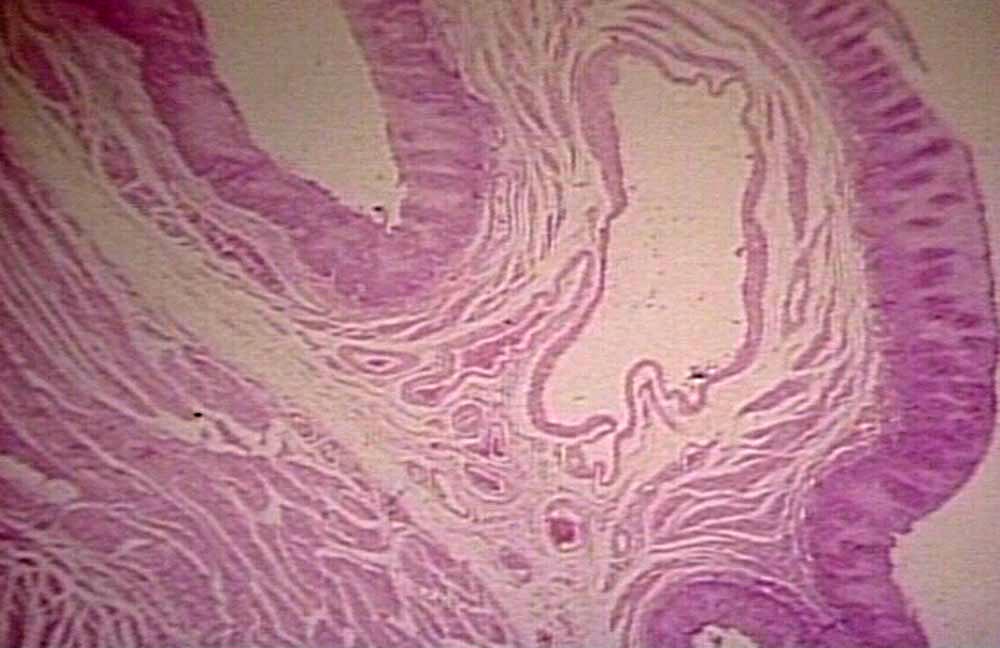
Microscopic
Features:
- Large dilated submucosal veins - key feature.
- +/-Blood (fresh).
- +/-Hemosiderin-laden macrophages.
Image:
See also
References
- ↑ Tsochatzis, EA.; Triantos, CK.; Garcovich, M.; Burroughs, AK. (Feb 2011). "Primary prevention of variceal hemorrhage.". Curr Gastroenterol Rep 13 (1): 3-9. doi:10.1007/s11894-010-0160-x. PMID 21086193.
- ↑ Burton, Julian L.; Rutty, Guy N. (2010). The Hospital Autopsy A Manual of Fundamental Autopsy Practice (3rd ed.). Oxford University Press. pp. 140. ISBN 978-0340965146.
- ↑ URL: http://www.pathguy.com/lectures/guts.htm. Accessed on: 24 April 2013.