Difference between revisions of "Epithelioid hemangioendothelioma"
Jump to navigation
Jump to search
(redirect for now) |
|||
| (21 intermediate revisions by the same user not shown) | |||
| Line 1: | Line 1: | ||
{{ Infobox diagnosis | |||
| Name = {{PAGENAME}} | |||
| Image = Epithelioid_hemangioendothelioma.jpg | |||
| Width = | |||
| Caption = Epithelioid hemangioendothelioma. [[H&E stain]]. | |||
| Synonyms = | |||
| Micro = large epithelioid perivascular cells with abundant pale eosinophilic cytoplasm and cytoplasmic vacuolation ("blister cells") - may form lumen and have RBC within, vesicular nucleus +/-prominent nucleolus; tuft-like projections into capillaries; cells may be in well-circumscribed paucicellular nodules ''or'' poorly formed cellular aggregates | |||
| Subtypes = | |||
| LMDDx = epithelioid [[angiosarcoma]], [[hemangioma]], [[epithelioid sarcoma]] | |||
| Stains = | |||
| IHC = CD31 +ve, CD34 +ve, factor VIII +ve, CAMTA1 +ve, TFE3 +ve/-ve | |||
| EM = | |||
| Molecular = gene fusions: WWTR1-CAMTA1 (approximately 90% of cases), YAP1-TFE3 (small number of cases) | |||
| IF = | |||
| Gross = | |||
| Grossing = | |||
| Site = [[soft tissue lesions|soft tissue]] - see ''[[vascular tumours]]'', classically [[liver]] - but various sites reported | |||
| Assdx = | |||
| Syndromes = | |||
| Clinicalhx = | |||
| Signs = | |||
| Symptoms = | |||
| Prevalence = rare | |||
| Bloodwork = | |||
| Rads = | |||
| Endoscopy = | |||
| Prognosis = moderate | |||
| Other = | |||
| ClinDDx = | |||
| Tx = resection | |||
}} | |||
'''Epithelioid hemangioendothelioma''', abbreviated '''EHE''', is rare malignant [[vascular tumours|vascular tumour]]. | |||
It should '''not''' be confused with ''[[epithelioid hemangioma]]''. | |||
==General== | |||
*Malignant.<ref name=Ref_WMSP603>{{Ref WMSP|603}}</ref> | |||
*Adults - wide age range. | |||
*Associated with [[oral contraceptives]], vinyl chloride.<ref name=pmid20165548/> | |||
*Rare.<ref name=pmid23589078/> | |||
Treatment: | |||
*Excision<ref name=pmid8941001/> if feasible. | |||
*Chemotherapy - not standardized.<ref name=pmid23589078>{{Cite journal | last1 = Chevreau | first1 = C. | last2 = Le Cesne | first2 = A. | last3 = Ray-Coquard | first3 = I. | last4 = Italiano | first4 = A. | last5 = Cioffi | first5 = A. | last6 = Isambert | first6 = N. | last7 = Robin | first7 = YM. | last8 = Fournier | first8 = C. | last9 = Clisant | first9 = S. | title = Sorafenib in patients with progressive epithelioid hemangioendothelioma: a phase 2 study by the French Sarcoma Group (GSF/GETO). | journal = Cancer | volume = 119 | issue = 14 | pages = 2639-44 | month = Jul | year = 2013 | doi = 10.1002/cncr.28109 | PMID = 23589078 }}</ref> | |||
*[[Liver transplantation]].<ref>{{Cite journal | last1 = Nudo | first1 = CG. | last2 = Yoshida | first2 = EM. | last3 = Bain | first3 = VG. | last4 = Marleau | first4 = D. | last5 = Wong | first5 = P. | last6 = Marotta | first6 = PJ. | last7 = Renner | first7 = E. | last8 = Watt | first8 = KD. | last9 = Deschênes | first9 = M. | title = Liver transplantation for hepatic epithelioid hemangioendothelioma: the Canadian multicentre experience. | journal = Can J Gastroenterol | volume = 22 | issue = 10 | pages = 821-4 | month = Oct | year = 2008 | doi = | PMID = 18925305 }}</ref> | |||
Prognosis - liver: | |||
*~55% five-year survival.<ref name=pmid8941001>{{Cite journal | last1 = Läuffer | first1 = JM. | last2 = Zimmermann | first2 = A. | last3 = Krähenbühl | first3 = L. | last4 = Triller | first4 = J. | last5 = Baer | first5 = HU. | title = Epithelioid hemangioendothelioma of the liver. A rare hepatic tumor. | journal = Cancer | volume = 78 | issue = 11 | pages = 2318-27 | month = Dec | year = 1996 | doi = | PMID = 8941001 }}</ref> | |||
**Better than other [[liver tumours]]. | |||
==Gross== | |||
*Classically, a [[liver]] lesion - but found elsewhere.<ref>{{Cite journal | last1 = Cardinal | first1 = J. | last2 = de Vera | first2 = ME. | last3 = Marsh | first3 = JW. | last4 = Steel | first4 = JL. | last5 = Geller | first5 = DA. | last6 = Fontes | first6 = P. | last7 = Nalesnik | first7 = M. | last8 = Gamblin | first8 = TC. | title = Treatment of hepatic epithelioid hemangioendothelioma: a single-institution experience with 25 cases. | journal = Arch Surg | volume = 144 | issue = 11 | pages = 1035-9 | month = Nov | year = 2009 | doi = 10.1001/archsurg.2009.121 | PMID = 19917940 }}</ref><ref name=pmid37541086>{{cite journal |authors=Haughey AM, Moloney BM, O'Brien CM |title=Epithelioid Haemangioendothelioma; Not simply a hepatic pathology |journal=Clin Imaging |volume=102 |issue= |pages=42–52 |date=October 2023 |pmid=37541086 |doi=10.1016/j.clinimag.2023.07.003 |url=}}</ref> | |||
*Case reports of EHE in a wide number of anatomical sites (bowel,<ref name=pmid30238810>{{cite journal |authors=Spasic S, Brcic I, Freire R, Garcia-Buitrago MT, Rosenberg AE |title=Epithelioid Hemangioendothelioma of the Bowel in Crohn's Disease: The First Reported Case |journal=Int J Surg Pathol |volume=27 |issue=4 |pages=423–426 |date=June 2019 |pmid=30238810 |doi=10.1177/1066896918801527 |url=}}</ref>, parotid<ref name=pmid31530411>{{cite journal |authors=Suarez-Zamora DA, Rodriguez-Urrego PA, Hakim-Tawil JA, Palau-Lazaro MA |title=Epithelioid hemangioendothelioma of the parotid gland: A case report in an unusual location with a review of the literature |journal=Rev Esp Patol |volume=52 |issue=4 |pages=260–264 |date=2019 |pmid=31530411 |doi=10.1016/j.patol.2019.04.002 |url=}}</ref> mediastinum<ref>{{cite journal |authors=Kim SH, Kim YS, Jang MH, Kwon HJ |title=Mediastinal Epithelioid Hemangioendothelioma Invading Superior Vena Cava: A Case Report and Review of Literature |journal=Curr Med Imaging Rev |volume=15 |issue=3 |pages=349–352 |date=2019 |pmid=31989887 |doi=10.2174/1573405614666180124141817 |url=}}</ref>). | |||
==Microscopic== | |||
Features:<ref name=pmid20165548>{{Cite journal | last1 = Gupta | first1 = R. | last2 = Mathur | first2 = SR. | last3 = Gupta | first3 = SD. | last4 = Durgapal | first4 = P. | last5 = Iyer | first5 = VK. | last6 = Das | first6 = CJ. | last7 = Shalimar | first7 = SK. | last8 = Acharya | first8 = . | title = Hepatic epithelioid hemangioendothelioma: A diagnostic pitfall in aspiration cytology. | journal = Cytojournal | volume = 6 | issue = | pages = 25 | month = | year = 2010 | doi = 10.4103/1742-6413.58951 | PMID = 20165548 | URL = http://www.biomedsearch.com/nih/Hepatic-epithelioid-hemangioendothelioma-diagnostic-pitfall/20165548.html }}</ref> | |||
*Large epithelioid perivascular cells with: | |||
**Abundant pale eosinophilic cytoplasm. | |||
**Cytoplasmic vacuolation (some cells) - [[AKA]] "blister cells" - '''key feature'''. | |||
***May form lumen and have RBC within. | |||
**Vesicular nucleus with prominent nucleolus in some cells. | |||
*Tuft-like projections into capillaries. | |||
*Tumour cells may be in well-circumscribed paucicellular nodules ''or'' more cellular poorly formed aggregates. | |||
DDx:<ref name=pmid19917940>{{Cite journal | last1 = Cardinal | first1 = J. | last2 = de Vera | first2 = ME. | last3 = Marsh | first3 = JW. | last4 = Steel | first4 = JL. | last5 = Geller | first5 = DA. | last6 = Fontes | first6 = P. | last7 = Nalesnik | first7 = M. | last8 = Gamblin | first8 = TC. | title = Treatment of hepatic epithelioid hemangioendothelioma: a single-institution experience with 25 cases. | journal = Arch Surg | volume = 144 | issue = 11 | pages = 1035-9 | month = Nov | year = 2009 | doi = 10.1001/archsurg.2009.121 | PMID = 19917940 }}</ref> | |||
*[[Angiosarcoma]], epithelioid. | |||
*[[Hemangioma]]. | |||
*[[Cholangiocarcinoma]]. | |||
*[[Fibrolamellar hepatocellular carcinoma]]. | |||
*[[Epithelioid sarcoma]].<ref name=pmid26414223/> | |||
===Images=== | |||
<gallery> | |||
Image:Epithelioid_hemangioendothelioma.jpg | Epithelioid hemangioendothelioma. (WC) | |||
</gallery> | |||
www: | |||
*[http://www.flickr.com/photos/pulmonary_pathology/5600772691/in/photostream/ Epithelioid hemangioendothelioma - low mag. (flickr.com/Rosen)]. | |||
*[http://www.flickr.com/photos/pulmonary_pathology/5601354962/ Epithelioid hemangioendothelioma - high mag. (flickr.com/Rosen)]. | |||
*[http://www.surgicalpathologyatlas.com/glfusion/mediagallery/media.php?f=0&sort=0&s=20080802170854495 Epithelioid hemangioendothelioma (surgicalpathologyatlas.com)]. | |||
==IHC== | |||
Features:<ref name=pmid20165548>{{Cite journal | last1 = Gupta | first1 = R. | last2 = Mathur | first2 = SR. | last3 = Gupta | first3 = SD. | last4 = Durgapal | first4 = P. | last5 = Iyer | first5 = VK. | last6 = Das | first6 = CJ. | last7 = Shalimar | first7 = SK. | last8 = Acharya | first8 = . | title = Hepatic epithelioid hemangioendothelioma: A diagnostic pitfall in aspiration cytology. | journal = Cytojournal | volume = 6 | issue = | pages = 25 | month = | year = 2010 | doi = 10.4103/1742-6413.58951 | PMID = 20165548 | URL = http://www.biomedsearch.com/nih/Hepatic-epithelioid-hemangioendothelioma-diagnostic-pitfall/20165548.html }}</ref> | |||
*CD31 +ve. | |||
*CD34 +ve. | |||
*Factor VIII +ve. | |||
*CAMTA1 +ve.<ref name=pmid26414223>{{cite journal |authors=Doyle LA, Fletcher CD, Hornick JL |title=Nuclear Expression of CAMTA1 Distinguishes Epithelioid Hemangioendothelioma From Histologic Mimics |journal=Am J Surg Pathol |volume=40 |issue=1 |pages=94–102 |date=January 2016 |pmid=26414223 |doi=10.1097/PAS.0000000000000511 |url=}}</ref> | |||
*TFE3 +ve - minority of cases. | |||
==Molecular== | |||
Gene fusions:<ref name=pmid26414223/> | |||
*WWTR1-CAMTA1 - seen in approximately 90% of cases. | |||
*YAP1-TFE3 fusion gene - <5% of cases. | |||
==See also== | |||
*[[Vascular tumours]]. | |||
*[[Kaposiform hemangioendothelioma]]. | |||
*[[Hemangioendothelioma]]. | |||
*[[Liver hemangioma]]. | |||
==References== | |||
{{Reflist|2}} | |||
[[Category:Diagnosis]] | |||
[[Category:Vascular tumours]] | |||
Latest revision as of 14:31, 5 April 2024
| Epithelioid hemangioendothelioma | |
|---|---|
| Diagnosis in short | |
 Epithelioid hemangioendothelioma. H&E stain. | |
|
| |
| LM | large epithelioid perivascular cells with abundant pale eosinophilic cytoplasm and cytoplasmic vacuolation ("blister cells") - may form lumen and have RBC within, vesicular nucleus +/-prominent nucleolus; tuft-like projections into capillaries; cells may be in well-circumscribed paucicellular nodules or poorly formed cellular aggregates |
| LM DDx | epithelioid angiosarcoma, hemangioma, epithelioid sarcoma |
| IHC | CD31 +ve, CD34 +ve, factor VIII +ve, CAMTA1 +ve, TFE3 +ve/-ve |
| Molecular | gene fusions: WWTR1-CAMTA1 (approximately 90% of cases), YAP1-TFE3 (small number of cases) |
| Site | soft tissue - see vascular tumours, classically liver - but various sites reported |
|
| |
| Prevalence | rare |
| Prognosis | moderate |
| Treatment | resection |
Epithelioid hemangioendothelioma, abbreviated EHE, is rare malignant vascular tumour.
It should not be confused with epithelioid hemangioma.
General
- Malignant.[1]
- Adults - wide age range.
- Associated with oral contraceptives, vinyl chloride.[2]
- Rare.[3]
Treatment:
- Excision[4] if feasible.
- Chemotherapy - not standardized.[3]
- Liver transplantation.[5]
Prognosis - liver:
- ~55% five-year survival.[4]
- Better than other liver tumours.
Gross
- Classically, a liver lesion - but found elsewhere.[6][7]
- Case reports of EHE in a wide number of anatomical sites (bowel,[8], parotid[9] mediastinum[10]).
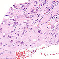
Microscopic
Features:[2]
- Large epithelioid perivascular cells with:
- Abundant pale eosinophilic cytoplasm.
- Cytoplasmic vacuolation (some cells) - AKA "blister cells" - key feature.
- May form lumen and have RBC within.
- Vesicular nucleus with prominent nucleolus in some cells.
- Tuft-like projections into capillaries.
- Tumour cells may be in well-circumscribed paucicellular nodules or more cellular poorly formed aggregates.
DDx:[11]
- Angiosarcoma, epithelioid.
- Hemangioma.
- Cholangiocarcinoma.
- Fibrolamellar hepatocellular carcinoma.
- Epithelioid sarcoma.[12]
Images
Epithelioid hemangioendothelioma. (WC)
www:
- Epithelioid hemangioendothelioma - low mag. (flickr.com/Rosen).
- Epithelioid hemangioendothelioma - high mag. (flickr.com/Rosen).
- Epithelioid hemangioendothelioma (surgicalpathologyatlas.com).
IHC
Features:[2]
- CD31 +ve.
- CD34 +ve.
- Factor VIII +ve.
- CAMTA1 +ve.[12]
- TFE3 +ve - minority of cases.
Molecular
Gene fusions:[12]
- WWTR1-CAMTA1 - seen in approximately 90% of cases.
- YAP1-TFE3 fusion gene - <5% of cases.
See also
References
- ↑ Humphrey, Peter A; Dehner, Louis P; Pfeifer, John D (2008). The Washington Manual of Surgical Pathology (1st ed.). Lippincott Williams & Wilkins. pp. 603. ISBN 978-0781765275.
- ↑ 2.0 2.1 2.2 Gupta, R.; Mathur, SR.; Gupta, SD.; Durgapal, P.; Iyer, VK.; Das, CJ.; Shalimar, SK.; Acharya, . (2010). "Hepatic epithelioid hemangioendothelioma: A diagnostic pitfall in aspiration cytology.". Cytojournal 6: 25. doi:10.4103/1742-6413.58951. PMID 20165548.
- ↑ 3.0 3.1 Chevreau, C.; Le Cesne, A.; Ray-Coquard, I.; Italiano, A.; Cioffi, A.; Isambert, N.; Robin, YM.; Fournier, C. et al. (Jul 2013). "Sorafenib in patients with progressive epithelioid hemangioendothelioma: a phase 2 study by the French Sarcoma Group (GSF/GETO).". Cancer 119 (14): 2639-44. doi:10.1002/cncr.28109. PMID 23589078.
- ↑ 4.0 4.1 Läuffer, JM.; Zimmermann, A.; Krähenbühl, L.; Triller, J.; Baer, HU. (Dec 1996). "Epithelioid hemangioendothelioma of the liver. A rare hepatic tumor.". Cancer 78 (11): 2318-27. PMID 8941001.
- ↑ Nudo, CG.; Yoshida, EM.; Bain, VG.; Marleau, D.; Wong, P.; Marotta, PJ.; Renner, E.; Watt, KD. et al. (Oct 2008). "Liver transplantation for hepatic epithelioid hemangioendothelioma: the Canadian multicentre experience.". Can J Gastroenterol 22 (10): 821-4. PMID 18925305.
- ↑ Cardinal, J.; de Vera, ME.; Marsh, JW.; Steel, JL.; Geller, DA.; Fontes, P.; Nalesnik, M.; Gamblin, TC. (Nov 2009). "Treatment of hepatic epithelioid hemangioendothelioma: a single-institution experience with 25 cases.". Arch Surg 144 (11): 1035-9. doi:10.1001/archsurg.2009.121. PMID 19917940.
- ↑ Haughey AM, Moloney BM, O'Brien CM (October 2023). "Epithelioid Haemangioendothelioma; Not simply a hepatic pathology". Clin Imaging 102: 42–52. doi:10.1016/j.clinimag.2023.07.003. PMID 37541086.
- ↑ Spasic S, Brcic I, Freire R, Garcia-Buitrago MT, Rosenberg AE (June 2019). "Epithelioid Hemangioendothelioma of the Bowel in Crohn's Disease: The First Reported Case". Int J Surg Pathol 27 (4): 423–426. doi:10.1177/1066896918801527. PMID 30238810.
- ↑ Suarez-Zamora DA, Rodriguez-Urrego PA, Hakim-Tawil JA, Palau-Lazaro MA (2019). "Epithelioid hemangioendothelioma of the parotid gland: A case report in an unusual location with a review of the literature". Rev Esp Patol 52 (4): 260–264. doi:10.1016/j.patol.2019.04.002. PMID 31530411.
- ↑ Kim SH, Kim YS, Jang MH, Kwon HJ (2019). "Mediastinal Epithelioid Hemangioendothelioma Invading Superior Vena Cava: A Case Report and Review of Literature". Curr Med Imaging Rev 15 (3): 349–352. doi:10.2174/1573405614666180124141817. PMID 31989887.
- ↑ Cardinal, J.; de Vera, ME.; Marsh, JW.; Steel, JL.; Geller, DA.; Fontes, P.; Nalesnik, M.; Gamblin, TC. (Nov 2009). "Treatment of hepatic epithelioid hemangioendothelioma: a single-institution experience with 25 cases.". Arch Surg 144 (11): 1035-9. doi:10.1001/archsurg.2009.121. PMID 19917940.
- ↑ 12.0 12.1 12.2 Doyle LA, Fletcher CD, Hornick JL (January 2016). "Nuclear Expression of CAMTA1 Distinguishes Epithelioid Hemangioendothelioma From Histologic Mimics". Am J Surg Pathol 40 (1): 94–102. doi:10.1097/PAS.0000000000000511. PMID 26414223.