Difference between revisions of "Emphysema"
Jump to navigation
Jump to search
| (5 intermediate revisions by the same user not shown) | |||
| Line 19: | Line 19: | ||
| Syndromes = [[Alpha-1 antitrypsin deficiency]], others | | Syndromes = [[Alpha-1 antitrypsin deficiency]], others | ||
| Clinicalhx = +/-[[smoking]] | | Clinicalhx = +/-[[smoking]] | ||
| Signs = barrel chest | | Signs = barrel-shaped chest | ||
| Symptoms = shortness of breath | | Symptoms = shortness of breath | ||
| Prevalence = common | | Prevalence = common | ||
| Bloodwork = | | Bloodwork = | ||
| Rads = hyperinflation | | Rads = hyperinflation, Saber-sheath trachea (associated with COPD) | ||
| Endoscopy = | | Endoscopy = | ||
| Prognosis = dependent on underlying cause | | Prognosis = dependent on underlying cause | ||
| Line 64: | Line 64: | ||
*Bleb = (small) [[vesicle]].<ref>URL: [http://dictionary.reference.com/browse/bleb http://dictionary.reference.com/browse/bleb]. Accessed on: 3 August 2011.</ref> | *Bleb = (small) [[vesicle]].<ref>URL: [http://dictionary.reference.com/browse/bleb http://dictionary.reference.com/browse/bleb]. Accessed on: 3 August 2011.</ref> | ||
*Bulla = large vesicle.<ref>URL: [http://dictionary.reference.com/browse/bulla http://dictionary.reference.com/browse/bulla]. Accessed on: 3 August 2011.</ref> | *Bulla = large vesicle.<ref>URL: [http://dictionary.reference.com/browse/bulla http://dictionary.reference.com/browse/bulla]. Accessed on: 3 August 2011.</ref> | ||
===Images=== | |||
<gallery> | |||
Image:Centrilobular emphysema 865 lores.jpg |Centrilobular emphysema. (WC/Edwin Ewing Jr.) | |||
</gallery> | |||
===Radiology=== | |||
*''Saber-sheath trachea'' - a finding associated with ''COPD''.<ref name=pmid26912770>{{cite journal |authors=Tunsupon P, Dhillon SS, Harris K, Alraiyes AH |title=Saber-sheath trachea in a patient with severe COPD |journal=BMJ Case Rep |volume=2016 |issue= |pages= |date=February 2016 |pmid=26912770 |pmc=4769447 |doi=10.1136/bcr-2016-214648 |url=}}</ref> | |||
**Trachea's anterior to posterior dimension:left to right dimension is >2:1.<ref>{{cite journal |authors=Webb EM, Elicker BM, Webb WR |title=Using CT to diagnose nonneoplastic tracheal abnormalities: appearance of the tracheal wall |journal=AJR Am J Roentgenol |volume=174 |issue=5 |pages=1315–21 |date=May 2000 |pmid=10789785 |doi=10.2214/ajr.174.5.1741315 |url=}}</ref> | |||
*Barrel-shaped chest.<ref name=pmid30604704>{{cite journal |vauthors=Sarkar M, Bhardwaz R, Madabhavi I, Modi M |title=Physical signs in patients with chronic obstructive pulmonary disease |journal=Lung India |volume=36 |issue=1 |pages=38–47 |date=2019 |pmid=30604704 |pmc=6330798 |doi=10.4103/lungindia.lungindia_145_18 |url=}}</ref> | |||
==Microscopic== | ==Microscopic== | ||
Latest revision as of 15:18, 24 November 2021
| Emphysema | |
|---|---|
| Diagnosis in short | |
 Emphysematous changes. H&E stain. | |
|
| |
| LM | alveoli too large, thin septa (no interstitial thickening) |
| Subtypes | centriacinar (centrilobular) emphysema, panacinar (panlobular) emphysema, distal (paraseptal) acinar emphysema, irregular emphysema |
| Gross | usually upper lobe predominant - blebs, bullae |
| Site | lung |
|
| |
| Associated Dx | +/-pneumothorax |
| Syndromes | Alpha-1 antitrypsin deficiency, others |
|
| |
| Clinical history | +/-smoking |
| Signs | barrel-shaped chest |
| Symptoms | shortness of breath |
| Prevalence | common |
| Radiology | hyperinflation, Saber-sheath trachea (associated with COPD) |
| Prognosis | dependent on underlying cause |
| Treatment | stop smoking, bullectomy |
Emphysema is a common medical lung disease strongly associated with smoking.
Chronic obstructive pulmonary disease, abbreviated COPD, redirects here.
General
- Usually due to smoking.
- Often lumped together with chronic bronchitis and called chronic obstructive pulmonary disease (COPD).[1]
- May cause pneumothorax - especially in young adults.[2]
Causes of emphysema other than smoking:[3]
Pathologic classification
Based on morphology:[4]
- Centriacinar (centrilobular) emphysema - associated with heavy smoking.
- Panacinar (panlobular) emphysema - associated with alpha-1 antitrypsin deficiency.
- Distal (paraseptal) acinar emphysema - associated with spontaneous pneumothorax.
- Irregular emphysema - usu. insignificant.
Note:
- Why does smoking lead to centriacinar emphysema?
- The bad stuff from smoking gets enters the acinus at the centre; ergo, this is the location of the most damage.
Gross
- Holes (blebs, bullae), usually upper lung field predominant.
- Lungs may overlap the heart.[5]
Notes:
Images
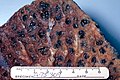
Centrilobular emphysema. (WC/Edwin Ewing Jr.)

Radiology
- Saber-sheath trachea - a finding associated with COPD.[8]
- Trachea's anterior to posterior dimension:left to right dimension is >2:1.[9]
- Barrel-shaped chest.[10]
Microscopic
Features:[5]
- Large alveoli.
- Thin septa (no interstitial thickening).
Images
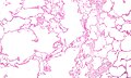
Emphysema. (WC)
See also
References
- ↑ Mitchell, Richard; Kumar, Vinay; Fausto, Nelson; Abbas, Abul K.; Aster, Jon (2011). Pocket Companion to Robbins & Cotran Pathologic Basis of Disease (8th ed.). Elsevier Saunders. pp. 368. ISBN 978-1416054542.
- ↑ Leslie, Kevin O.; Wick, Mark R. (2004). Practical Pulmonary Pathology: A Diagnostic Approach (1st ed.). Churchill Livingstone. pp. 296. ISBN 978-0443066313.
- ↑ Lee, P.; Gildea, TR.; Stoller, JK. (Dec 2002). "Emphysema in nonsmokers: alpha 1-antitrypsin deficiency and other causes.". Cleve Clin J Med 69 (12): 928-9, 933, 936 passim. PMID 12546267.
- ↑ Mitchell, Richard; Kumar, Vinay; Fausto, Nelson; Abbas, Abul K.; Aster, Jon (2011). Pocket Companion to Robbins & Cotran Pathologic Basis of Disease (8th ed.). Elsevier Saunders. pp. 368. ISBN 978-1416054542.
- ↑ 5.0 5.1 Mitchell, Richard; Kumar, Vinay; Fausto, Nelson; Abbas, Abul K.; Aster, Jon (2011). Pocket Companion to Robbins & Cotran Pathologic Basis of Disease (8th ed.). Elsevier Saunders. pp. 369. ISBN 978-1416054542.
- ↑ URL: http://dictionary.reference.com/browse/bleb. Accessed on: 3 August 2011.
- ↑ URL: http://dictionary.reference.com/browse/bulla. Accessed on: 3 August 2011.
- ↑ Tunsupon P, Dhillon SS, Harris K, Alraiyes AH (February 2016). "Saber-sheath trachea in a patient with severe COPD". BMJ Case Rep 2016. doi:10.1136/bcr-2016-214648. PMC 4769447. PMID 26912770. https://www.ncbi.nlm.nih.gov/pmc/articles/PMC4769447/.
- ↑ Webb EM, Elicker BM, Webb WR (May 2000). "Using CT to diagnose nonneoplastic tracheal abnormalities: appearance of the tracheal wall". AJR Am J Roentgenol 174 (5): 1315–21. doi:10.2214/ajr.174.5.1741315. PMID 10789785.
- ↑ "Physical signs in patients with chronic obstructive pulmonary disease". Lung India 36 (1): 38–47. 2019. doi:10.4103/lungindia.lungindia_145_18. PMC 6330798. PMID 30604704. https://www.ncbi.nlm.nih.gov/pmc/articles/PMC6330798/.