Dermal cysts
Jump to navigation
Jump to search
The printable version is no longer supported and may have rendering errors. Please update your browser bookmarks and please use the default browser print function instead.
Dermal cysts, also skin cysts, are common in dermatopathology. Dermatopathologists can diagnose 'em.
Overview
Common types:[1]
- Epidermal cyst (sebaceous cyst) -- most common.
- Pilar (trichilemmal) cyst.
- Dermoid cyst.
- Ganglion cyst.
- Milicem.
Epidermal necrosis
- This may be cystic. It is covered in the epidermal necrosis article, which covers erythema multiforme, Steven-Johnson syndrome and toxic epidermal necrolysis.
Common cysts
Venous lake
Main article: Venous lake
Epidermal inclusion cyst
Main article: Epidermal inclusion cyst
Pilar cyst
- AKA trichilemmal cyst.
Main article: Pilar cyst
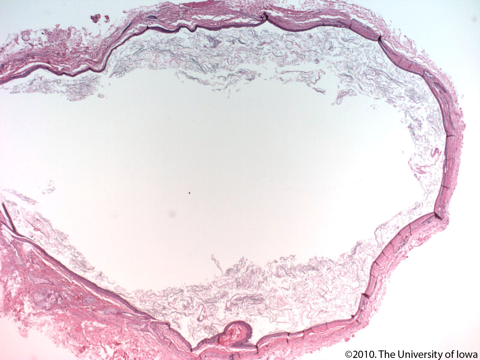
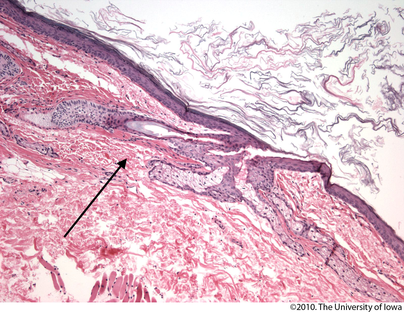
Dermoid cyst
General
- Benign.
- Congenital choristomas.[2]
- May be found in the ovary.
Microscopic
- Cyst lined by normal (keratinized) skin with adnexal structure (hair follicles, sweat glands, sebaceous glands).
DDx:
- Epidermal cyst - no adnexal structures.
Images:
Sign out
SKIN CYST, RIGHT LATERAL ORBIT, EXCISION:
- DERMOID CYST
- NEGATIVE FOR MALIGNANCY.
Pilonidal cyst
Main article: Pilonidal sinus
Less common
Steatocystoma
Main article: Steatocystoma
Digital mucous cyst
General
- Dome-shaped papule.
Microscopic
Features:[5]
- Mucous in superficial dermis - key feature.
- No epithelial lining; it is a pseudocyst.
Note:
- Mucin = glycolated proteins; may be part of mucous.
- Mucous = slippery secretion.
DDx:
Images:
Sign out
LESION, LEFT INDEX FINGER, EXCISION:
- DIGITAL MUCOUS CYST.
Apocrine cystadenoma
General
- Uncommon.
Microscopic
Features:[9]
- Multiloculated.
- Apocrine differentiation: columnar epithelium +/- apical snouts.
- Solid areas of epithelial proliferation.
- Papillary projections into the cyst.
Images:
See also
References
- ↑ Greenwald, J.; Heng, M. (2007). Toronto Notes for Medical Students 2007 (2007 ed.). The Toronto Notes Inc. for Medical Students Inc.. pp. D5. ISBN 978-0968592878.
- ↑ 2.0 2.1 2.2 Gandhi N, Syed NA, Alen R. Dermoid Cyst. EyeRounds.org. posted July 26, 2010; Available from: http://www.EyeRounds.org/cases/115-dermoid-cyst.htm. Accessed on: 22 September 2011.
- ↑ Mitchell, Richard; Kumar, Vinay; Fausto, Nelson; Abbas, Abul K.; Aster, Jon (2011). Pocket Companion to Robbins & Cotran Pathologic Basis of Disease (8th ed.). Elsevier Saunders. pp. 596. ISBN 978-1416054542.
- ↑ URL: http://emedicine.medscape.com/article/788127-overview. Accessed on: 10 September 2012.
- ↑ 5.0 5.1 5.2 5.3 URL: http://www.dermpedia.org/dermpedia-textbook/digital-mucous-myxoid-cyst. Accessed on: 17 January 2012.
- ↑ URL: http://dictionary.reference.com/browse/mucous. Accessed on: 8 January 2012.
- ↑ URL: http://dictionary.reference.com/browse/mucus. Accessed on: 8 January 2012.
- ↑ URL: http://www.dermpedia.org/case/digital-mucous-cyst-ganglion-type. Accessed on: 5 July 2013.
- ↑ Busam, Klaus J. (2009). Dermatopathology: A Volume in the Foundations in Diagnostic Pathology Series (1st ed.). Saunders. pp. 316. ISBN 978-0443066542.