Difference between revisions of "Crystals in body fluids"
Jump to navigation
Jump to search
| Line 53: | Line 53: | ||
=External links= | =External links= | ||
*[http://granuloma.homestead.com/foreignbody2.html Foreign body granulomas (granuloma.homestead.com)]. | *[http://granuloma.homestead.com/foreignbody2.html Foreign body granulomas (granuloma.homestead.com)]. | ||
*[http://www.eclinpath.com/atlas/urinalysis/urine-crystals/nggallery/page/3 Urine cytology -veterinary medicine (eclinpath.com)]. | *[http://www.eclinpath.com/atlas/urinalysis/urine-crystals/nggallery/page/3 Urine cytology - veterinary medicine (eclinpath.com)]. | ||
[[Category:Clinical]] | [[Category:Clinical]] | ||
Revision as of 03:24, 3 January 2016
.jpg)
This article deals with crystals in body fluids.
Crystals
Joint crystals
Types:[1]
- Gout = needle-shaped, negatively birefringent, yellow when aligned.
- Pseudogout = rhomboid-shaped, positively birefringent, blue when aligned.
Notes:
- Pseudogout also known as CPPD = calcium pyrophosphate dehydrogenase.
- Memory device: ABC+ = aligned blue is calcium & cuboid - positively birefringent.
Urine crystals
Types - morphology:
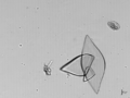
- Envelope shape (calcium oxalate).
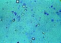
- Diamond shape (uric acid).
- Coffin-lid shape (struvite).
- Hexagonal shape (cysteine).
Notes:
- Memory devices:
- Diamonds are see-through; ergo, uric acid stones not seen on KUB.
- Calcium oxalate = envelope, uric acid = diamond.
- Uric acid crystals: usually dissolve in formalin... but do not dissolve in alcohol.[2][3]
- Calcium oxalate crystals are seen in the context of ethylene glycol poisoning.[4]
Images
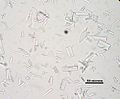
Struvite crystals. (WC)
Uric acid crystals. (WC)
Calcium oxalate crystal - envelope-shaped. (WC)

_-_%C3%9Crik_asit_kristalleri_(idrar)_-_03.png)

www:
Diseases
Gout
Main article: Gout
Pseudogout
Main article: Chondrocalcinosis
See also
- Cytopathology.
- Medical renal diseases.
- Nephrolithiasis - kidney stones.
References
- ↑ Yeung, J.C.; Leonard, Blair J. N. (2005). The Toronto Notes 2005 - Review for the MCCQE and Comprehensive Medical Reference (2005 ed.). The Toronto Notes Inc. for Medical Students Inc.. pp. RH6. ISBN 978-0968592854.
- ↑ Geddie, W. 8 January 2010.
- ↑ Shidham, V.; Chivukula, M.; Basir, Z.; Shidham, G. (Aug 2001). "Evaluation of crystals in formalin-fixed, paraffin-embedded tissue sections for the differential diagnosis of pseudogout, gout, and tumoral calcinosis.". Mod Pathol 14 (8): 806-10. doi:10.1038/modpathol.3880394. PMID 11504841.
- ↑ Saukko, Pekka; Knight, Bernard (2004). Knight's Forensic Pathology (3rd ed.). A Hodder Arnold Publication. pp. 589. ISBN 978-0340760444.
- ↑ URL: http://www.ncbi.nlm.nih.gov/pubmedhealth/PMH0001458/. Accessed on: 28 October 2011.