Difference between revisions of "Craniopharyngioma"
(→Papillary: replace ref, fix typo, tweak format) |
Jensflorian (talk | contribs) (histo + pictures added) |
||
| Line 67: | Line 67: | ||
==General== | ==General== | ||
*Develop from remains of Rathke's pouch or squamous epithelial cell rests.<ref name=pmid17425791>{{Cite journal | last1 = Garnett | first1 = MR. | last2 = Puget | first2 = S. | last3 = Grill | first3 = J. | last4 = Sainte-Rose | first4 = C. | title = Craniopharyngioma. | journal = Orphanet J Rare Dis | volume = 2 | issue = | pages = 18 | month = | year = 2007 | doi = 10.1186/1750-1172-2-18 | PMID = 17425791 }}</ref> | *Develop from remains of Rathke's pouch or squamous epithelial cell rests.<ref name=pmid17425791>{{Cite journal | last1 = Garnett | first1 = MR. | last2 = Puget | first2 = S. | last3 = Grill | first3 = J. | last4 = Sainte-Rose | first4 = C. | title = Craniopharyngioma. | journal = Orphanet J Rare Dis | volume = 2 | issue = | pages = 18 | month = | year = 2007 | doi = 10.1186/1750-1172-2-18 | PMID = 17425791 }}</ref> | ||
*corresponds histologically to WHO grade I. | |||
Subtypes:<ref name=pmid17425791/> | Subtypes:<ref name=pmid17425791/> | ||
| Line 89: | Line 90: | ||
*Calcified - adamantinomatous type only. | *Calcified - adamantinomatous type only. | ||
*Solid & cystic. | *Solid & cystic. | ||
====Images==== | |||
<gallery> | |||
File:Craniopharyngioma1.jpg|Calcifications in adamantinomatous craniopharyngioma. (WC/Garnett et al.) | |||
File:Papillary craniopharyngioma.jpg|Autopsy case with papillary craniopharyngioma of the 3rd ventricle. (WC/AFIP) | |||
File:Adamantinomatous craniopharyngioma.jpg|Cholesterol crystals, a typical finding in the cyst fluid, are readily identified by examination under polarized light. (WC/AFIP) | |||
</gallery> | |||
==Microscopic== | ==Microscopic== | ||
===Adamantinomatous=== | ===Adamantinomatous=== | ||
Features (adamantinomatous):<ref name=Ref_DCHH184>{{Ref DCHH|184}}</ref> | Features (adamantinomatous):<ref name=Ref_DCHH184>{{Ref DCHH|184}}</ref> | ||
*trabecular squamous epithelium bordered by palisaded columar epithelum. | |||
*Lobules with loosely distributed epithelia ("stellate reticulum"). | |||
*Well-circumscribed (or pseudoinvasive border). | *Well-circumscribed (or pseudoinvasive border). | ||
*Multicystic. | *Multicystic. | ||
| Line 114: | Line 124: | ||
Image:CNS Craniopharyngeoma Adamantinomatous MP MP PA.JPG|Adamantinomatous Craniopharyngeoma - medium power (SKB) | Image:CNS Craniopharyngeoma Adamantinomatous MP MP PA.JPG|Adamantinomatous Craniopharyngeoma - medium power (SKB) | ||
Image:CNS Craniopharyngeoma Adamantinomatous 2 MP PA.JPG|Adamantinomatous Craniopharyngeoma - medium power (SKB) | Image:CNS Craniopharyngeoma Adamantinomatous 2 MP PA.JPG|Adamantinomatous Craniopharyngeoma - medium power (SKB) | ||
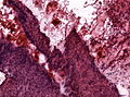
Image:Craniopharyngeoma CNS infiltration.jpg|Brain infiltration. "Wet" keratin present in two tumor protrusions. The surrounding CNS shows extensive piloid gliosis. (WC/jensflorian) | |||
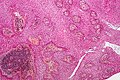
Image:Craniopharyngioma histology.jpg|Adamantinomatous Craniopharyngeoma - Trabecular growth pattern. (WC/jensflorian) | |||
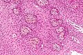
Image:Craniopharyngioma_HE_frozen.jpg|Adamantinomatous Craniopharyngeoma - Intraoperative frozen section. (WC/jensflorian) | |||
</gallery> | </gallery> | ||
| Line 125: | Line 138: | ||
*+/-Goblet cell-like formations (rare). | *+/-Goblet cell-like formations (rare). | ||
==== | ====Images==== | ||
<gallery> | <gallery> | ||
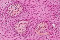
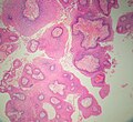
Image: Papillary craniopharyngioma - intermed mag.jpg | Papillary craniopharyngioma - intermed. mag. (WC/Nephron) | Image: Papillary craniopharyngioma - intermed mag.jpg | Papillary craniopharyngioma - intermed. mag. (WC/Nephron) | ||
| Line 140: | Line 153: | ||
*[http://www.cnsatlas.com/cgi-bin/nephrology/preview?ADD=0&LESION_ID=382&BOOK_ID=5&POST=toc Craniopharyngioma at cnsatlas.com].<ref>URL:[http://www.cnsatlas.com/cgi-bin/nephrology/preview?ADD=0&LESION_ID=382&BOOK_ID=5&POST=toc]. Accessed on: 21 March 2015.</ref> | *[http://www.cnsatlas.com/cgi-bin/nephrology/preview?ADD=0&LESION_ID=382&BOOK_ID=5&POST=toc Craniopharyngioma at cnsatlas.com].<ref>URL:[http://www.cnsatlas.com/cgi-bin/nephrology/preview?ADD=0&LESION_ID=382&BOOK_ID=5&POST=toc]. Accessed on: 21 March 2015.</ref> | ||
*[http://neuropathology-web.org/chapter7/chapter7dMiscellaneous.html Craniopharyngioma at Neuropathology-web.org]<ref>URL:http://neuropathology-web.org/chapter7/chapter7dMiscellaneous.html]. Accessed on: 21 March 2015.</ref> | *[http://neuropathology-web.org/chapter7/chapter7dMiscellaneous.html Craniopharyngioma at Neuropathology-web.org]<ref>URL:http://neuropathology-web.org/chapter7/chapter7dMiscellaneous.html]. Accessed on: 21 March 2015.</ref> | ||
===Differential diagnosis=== | |||
*Xanthogranuloma | |||
*Rathke cyst | |||
*Epidermoid | |||
*Well-differentiated carcinoma metastasis | |||
==See also== | ==See also== | ||
Revision as of 10:26, 30 March 2015
| Adamantinomatous craniopharyngioma | |
|---|---|
| Diagnosis in short | |
 Adamantinomatous craniopharyngioma. HPS stain. | |
|
| |
| LM | well-circumscribed (or pseudoinvasive border), multicystic, small-to-medium sized cells with moderate amount of basophilic cytoplasm, bland nuclei (with occ. small nucleoli), "wet" keratin (nests of whorled keratin), calcifications |
| Gross | cystic mass filled with motor oil-like fluid |
| Site | sella turcica |
|
| |
| Clinical history | adults & children |
| Radiology | classically calcified |
| Prognosis | benign |
| Clin. DDx | other sella turcica lesions |
| Papillary craniopharyngioma | |
|---|---|
| Diagnosis in short | |
 Papillary craniopharyngioma. HPS stain. | |
|
| |
| LM | non-keratinized squamous epithelium (without nuclear atypia), fibrovascular cores (required for papillary) |
| Site | sella turcica |
|
| |
| Clinical history | adults |
| Prognosis | benign |
| Clin. DDx | other sella turcica lesions |
Craniopharyngioma is a benign neuropathology tumour.
It is subdivided into papillary craniopharyngioma and adamantinomatous craniopharyngioma.
General
- Develop from remains of Rathke's pouch or squamous epithelial cell rests.[1]
- corresponds histologically to WHO grade I.
Subtypes:[1]
- Adamantinomatous type.
- Squamous papillary type.
Adamantinomatous
- Adults and children.
- Typically contain mutations in CTNNB1 (the gene that encodes β-catenin).[2]
- Usually intranuclear β-catenin immunohistochemical positivity.
Papillary
Gross
- Cystic mass filled with motor oil-like fluid.[5]
- May not be seen in the papillary variant of craniopharyngioma.
Radiology:[1]
- Calcified - adamantinomatous type only.
- Solid & cystic.
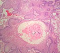
Images
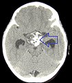
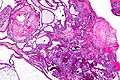
Calcifications in adamantinomatous craniopharyngioma. (WC/Garnett et al.)
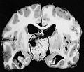
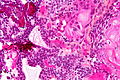
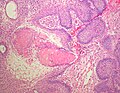
Autopsy case with papillary craniopharyngioma of the 3rd ventricle. (WC/AFIP)
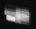
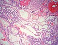
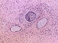
Cholesterol crystals, a typical finding in the cyst fluid, are readily identified by examination under polarized light. (WC/AFIP)


Microscopic
Adamantinomatous
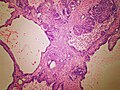
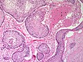
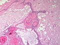
Features (adamantinomatous):[6]
- trabecular squamous epithelium bordered by palisaded columar epithelum.
- Lobules with loosely distributed epithelia ("stellate reticulum").
- Well-circumscribed (or pseudoinvasive border).
- Multicystic.
- Small-to-medium sized cells with moderate amount of basophilic cytoplasm.
- Bland nuclei (with occ. small nucleoli).
- "Wet" keratin - nests of whorled keratin.
- Calcifications (non-psammomatous).
Images
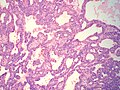
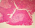
Adamantinomatous craniopharyngioma - very low mag. (WC/Nephron)
Adamantinomatous craniopharyngioma - intermed. mag. (WC/Nephron)
Adamantinomatous craniopharyngioma - very high mag. (WC/Nephron)
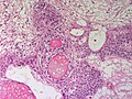
Adamantinomatous Craniopharyngeoma - low power (SKB)
Adamantinomatous Craniopharyngeoma - low power (SKB)
Adamantinomatous Craniopharyngeoma - low power (SKB)
Adamantinomatous Craniopharyngeoma - medium power (SKB)
Adamantinomatous Craniopharyngeoma - medium power (SKB)
Adamantinomatous Craniopharyngeoma - medium power (SKB)
Adamantinomatous Craniopharyngeoma - medium power (SKB)
Adamantinomatous Craniopharyngeoma - medium power (SKB)
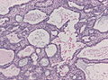
Brain infiltration. "Wet" keratin present in two tumor protrusions. The surrounding CNS shows extensive piloid gliosis. (WC/jensflorian)
Adamantinomatous Craniopharyngeoma - Trabecular growth pattern. (WC/jensflorian)
Adamantinomatous Craniopharyngeoma - Intraoperative frozen section. (WC/jensflorian)


Papillary
Features (papillary):[7]
- Non-keratinized squamous epithelium (without nuclear atypia).
- Fibrovascular cores (required for papillary).
Notes:
- +/-Cilia (rare).
- +/-Goblet cell-like formations (rare).
Images
Papillary craniopharyngioma - intermed. mag. (WC/Nephron)
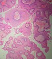
Papillary craniopharyngioma - high mag. (WC/Nephron)
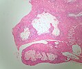
Papillary craniopharyngioma - very high mag. (WC/Nephron)
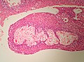
Papillary Craniopharyngeoma - low power (SKB)
Papillary Craniopharyngeoma - low power (SKB)
Papillary Craniopharyngeoma - low power (SKB)
Papillary Craniopharyngeoma - medium power (SKB)
Papillary Craniopharyngeoma - medium power (SKB)


_copy.jpg)


www:
- Craniopharyngioma (med.utah.edu).[8]
- Craniopharyngioma at cnsatlas.com.[9]
- Craniopharyngioma at Neuropathology-web.org[10]
Differential diagnosis
- Xanthogranuloma
- Rathke cyst
- Epidermoid
- Well-differentiated carcinoma metastasis
See also
References
- ↑ 1.0 1.1 1.2 Garnett, MR.; Puget, S.; Grill, J.; Sainte-Rose, C. (2007). "Craniopharyngioma.". Orphanet J Rare Dis 2: 18. doi:10.1186/1750-1172-2-18. PMID 17425791.
- ↑ Preda, V.; Larkin, SJ.; Karavitaki, N.; Ansorge, O.; Grossman, AB. (Oct 2014). "The Wnt Signalling Cascade and the Adherens Junction Complex in Craniopharyngioma Tumorigenesis.". Endocr Pathol. doi:10.1007/s12022-014-9341-8. PMID 25355426.
- ↑ Giangaspero, F.; Burger, PC.; Osborne, DR.; Stein, RB. (Jan 1984). "Suprasellar papillary squamous epithelioma ("papillary craniopharyngioma").". Am J Surg Pathol 8 (1): 57-64. PMID 6696166.
- ↑ Brastianos, PK.; Taylor-Weiner, A.; Manley, PE.; Jones, RT.; Dias-Santagata, D.; Thorner, AR.; Lawrence, MS.; Rodriguez, FJ. et al. (Feb 2014). "Exome sequencing identifies BRAF mutations in papillary craniopharyngiomas.". Nat Genet 46 (2): 161-5. doi:10.1038/ng.2868. PMID 24413733.
- ↑ Fernandez-Miranda, JC.; Gardner, PA.; Snyderman, CH.; Devaney, KO.; Strojan, P.; Suárez, C.; Genden, EM.; Rinaldo, A. et al. (Jul 2012). "Craniopharyngioma: a pathologic, clinical, and surgical review.". Head Neck 34 (7): 1036-44. doi:10.1002/hed.21771. PMID 21584897.
- ↑ Tadrous, Paul.J. Diagnostic Criteria Handbook in Histopathology: A Surgical Pathology Vade Mecum (1st ed.). Wiley. pp. 184. ISBN 978-0470519035.
- ↑ Perry, Arie; Brat, Daniel J. (2010). Practical Surgical Neuropathology: A Diagnostic Approach: A Volume in the Pattern Recognition series (1st ed.). Churchill Livingstone. pp. 406. ISBN 978-0443069826.
- ↑ URL: http://library.med.utah.edu/WebPath/jpeg4/ENDO115.jpg. Accessed on: 6 December 2010.
- ↑ URL:[1]. Accessed on: 21 March 2015.
- ↑ URL:http://neuropathology-web.org/chapter7/chapter7dMiscellaneous.html]. Accessed on: 21 March 2015.