Difference between revisions of "Condyloma acuminatum"
(+cat.) |
(split out) |
||
| Line 1: | Line 1: | ||
'''Condyloma acuminatum''', also '''genital wart''', is a common benign pathology of the genital region ([[vulva]], [[penis]], perineum). | |||
==General== | |||
*Due to [[human papillomavirus]] (HPV). | |||
**Transmission: sexual, non-sexual, horizontal (mother to child).<ref name=Ref_APBR280>{{Ref APBR|280 Q29}}</ref> | |||
***Should raise the suspicion of child abuse. | |||
Note: | |||
*Related to [[verruca vulgaris]] (common wart). | |||
*The Bethesda system includes this in [[LSIL]].<ref>{{Ref GP|143}}</ref> | |||
Clinical DDx: | |||
*[[Molluscum contagiosum]].<ref>URL: [http://emedicine.medscape.com/article/781735-differential http://emedicine.medscape.com/article/781735-differential]. Accessed on: 5 July 2013.</ref> | |||
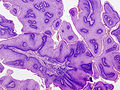
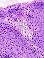
==Microscopic== | |||
Features: | |||
*Koilocytes.<ref name=pmid11860848>{{Cite journal | last1 = Huang | first1 = Z. | last2 = Yang | first2 = S. | last3 = Li | first3 = Q. | last4 = Yan | first4 = P. | last5 = Li | first5 = L. | title = [Evaluation the pathological diagnostic values of koilocyte in condyloma acuminatum]. | journal = Zhonghua Liu Xing Bing Xue Za Zhi | volume = 22 | issue = 1 | pages = 58-60 | month = Feb | year = 2001 | doi = | PMID = 11860848 }}</ref> | |||
**Cells with an enlarged nucleus and perinuclear clearing. | |||
*Papillomatosis.<ref>{{Ref WMSP|204}}</ref> | |||
**Papillomatosis = surface elevation due to dermal papillae enlargement.<ref>{{Ref PBoD|1230}}</ref> | |||
*+/-Parakeratosis. | |||
DDx: | |||
*[[Classic vulvar intraepithelial neoplasia]] - architecture different. | |||
===Images=== | |||
<gallery> | |||
Image:Condyloma_acuminatum_-_low_mag.jpg | Condyloma acuminatum - low mag. (WC) | |||
Image:Condyloma_acuminatum_-_very_high_mag.jpg | Condyloma acuminatum - very high mag. (WC) | |||
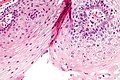
Image:Anal_condyloma_%282%29.jpg | Condyloma acuminatum - 2. (WC) | |||
Image:Anal_condyloma_%284%29.jpg | Condyloma acuminatum - 3. (WC) | |||
</gallery> | |||
==Sign out== | |||
<pre> | |||
SKIN LESION ("VULVAR WART"), VULVA, EXCISION: | |||
- CONDYLOMA ACUMINATUM (GENITAL WART). | |||
</pre> | |||
<pre> | |||
LABIA MINORA, BIOPSY: | |||
- CONDYLOMA/LOW-GRADE SQUAMOUS INTRAEPITHELIAL LESION (LSIL). | |||
-- NEGATIVE FOR HIGH-GRADE DYSPLASIA. | |||
</pre> | |||
===Seborrheic keratosis-like=== | |||
<pre> | |||
Submitted as "Penile Wart", Excision: | |||
- Consistent with condyloma acuminatum (genital wart) with | |||
seborrheic keratosis-like features. | |||
</pre> | |||
====Block letters==== | |||
<pre> | |||
SKIN LESION, PERINEUM, BIOPSY: | |||
- SEBORRHEIC KERATOSIS-LIKE CONDYLOMA ACUMINATUM (GENITAL WART). | |||
</pre> | |||
===Without viral cytopathic changes=== | |||
<pre> | |||
VULVAR LESIONS (x3), EXCISION: | |||
- SQUAMOUS HYPERPLASIA WITH HYPERORTHOKERATOSIS WITHOUT VIRAL CYTOPATHIC EFFECT, | |||
COMPATIBLE WITH CONDYLOMA (x3). | |||
- NEGATIVE FOR MALIGNANCY. | |||
</pre> | |||
===Micro=== | |||
The sections show a polypoid fragment of skin with epithelium on three sides, acanthosis, hyperkeratosis and parakeratosis. Koilocytic changes (mild nuclear enlargement, perinuclear clearing) are seen focally. There is mild basilar nuclear enlargement and hyperchromasia. The epithelium shows maturation to the surface and a granular layer is present. | |||
====Seborrheic keratosis-like==== | |||
The sections show skin with acanthosis with papillomatous features (round bulbous rete ridges, acanthosis with penetrating fibrovascular cores) pseudohorn cysts, parakeratosis and hyperkeratosis. There is no significant basal nuclear atypia. There are no mitoses and no melanocytic nests. There is mild dermal inflammation. There is no solar elastosis. Pigment incontinence is present focally. | |||
==See also== | |||
*[[Vulva]]. | |||
*[[Penis]]. | |||
==References== | |||
{{Reflist|2}} | |||
[[Category:Vulva]] | |||
[[Category:Diagnosis]] | [[Category:Diagnosis]] | ||
Revision as of 15:43, 8 March 2016
Condyloma acuminatum, also genital wart, is a common benign pathology of the genital region (vulva, penis, perineum).
General
- Due to human papillomavirus (HPV).
- Transmission: sexual, non-sexual, horizontal (mother to child).[1]
- Should raise the suspicion of child abuse.
- Transmission: sexual, non-sexual, horizontal (mother to child).[1]
Note:
- Related to verruca vulgaris (common wart).
- The Bethesda system includes this in LSIL.[2]
Clinical DDx:
Microscopic
Features:
- Koilocytes.[4]
- Cells with an enlarged nucleus and perinuclear clearing.
- Papillomatosis.[5]
- Papillomatosis = surface elevation due to dermal papillae enlargement.[6]
- +/-Parakeratosis.
DDx:
- Classic vulvar intraepithelial neoplasia - architecture different.
Images
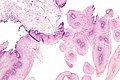
Condyloma acuminatum - low mag. (WC)
Condyloma acuminatum - very high mag. (WC)
Condyloma acuminatum - 2. (WC)
Condyloma acuminatum - 3. (WC)


.jpg)
.jpg)
Sign out
SKIN LESION ("VULVAR WART"), VULVA, EXCISION:
- CONDYLOMA ACUMINATUM (GENITAL WART).
LABIA MINORA, BIOPSY:
- CONDYLOMA/LOW-GRADE SQUAMOUS INTRAEPITHELIAL LESION (LSIL).
-- NEGATIVE FOR HIGH-GRADE DYSPLASIA.
Seborrheic keratosis-like
Submitted as "Penile Wart", Excision:
- Consistent with condyloma acuminatum (genital wart) with
  seborrheic keratosis-like features.
Block letters
SKIN LESION, PERINEUM, BIOPSY:
- SEBORRHEIC KERATOSIS-LIKE CONDYLOMA ACUMINATUM (GENITAL WART).
Without viral cytopathic changes
VULVAR LESIONS (x3), EXCISION:
- SQUAMOUS HYPERPLASIA WITH HYPERORTHOKERATOSIS WITHOUT VIRAL CYTOPATHIC EFFECT,
  COMPATIBLE WITH CONDYLOMA (x3).
- NEGATIVE FOR MALIGNANCY.
Micro
The sections show a polypoid fragment of skin with epithelium on three sides, acanthosis, hyperkeratosis and parakeratosis. Koilocytic changes (mild nuclear enlargement, perinuclear clearing) are seen focally. There is mild basilar nuclear enlargement and hyperchromasia. The epithelium shows maturation to the surface and a granular layer is present.
Seborrheic keratosis-like
The sections show skin with acanthosis with papillomatous features (round bulbous rete ridges, acanthosis with penetrating fibrovascular cores) pseudohorn cysts, parakeratosis and hyperkeratosis. There is no significant basal nuclear atypia. There are no mitoses and no melanocytic nests. There is mild dermal inflammation. There is no solar elastosis. Pigment incontinence is present focally.
See also
References
- ↑ Lefkowitch, Jay H. (2006). Anatomic Pathology Board Review (1st ed.). Saunders. pp. 280 Q29. ISBN 978-1416025887.
- ↑ Nucci, Marisa R.; Oliva, Esther (2009). Gynecologic Pathology: A Volume in Foundations in Diagnostic Pathology Series (1st ed.). Churchill Livingstone. pp. 143. ISBN 978-0443069208.
- ↑ URL: http://emedicine.medscape.com/article/781735-differential. Accessed on: 5 July 2013.
- ↑ Huang, Z.; Yang, S.; Li, Q.; Yan, P.; Li, L. (Feb 2001). "[Evaluation the pathological diagnostic values of koilocyte in condyloma acuminatum].". Zhonghua Liu Xing Bing Xue Za Zhi 22 (1): 58-60. PMID 11860848.
- ↑ Humphrey, Peter A; Dehner, Louis P; Pfeifer, John D (2008). The Washington Manual of Surgical Pathology (1st ed.). Lippincott Williams & Wilkins. pp. 204. ISBN 978-0781765275.
- ↑ Cotran, Ramzi S.; Kumar, Vinay; Fausto, Nelson; Nelso Fausto; Robbins, Stanley L.; Abbas, Abul K. (2005). Robbins and Cotran pathologic basis of disease (7th ed.). St. Louis, Mo: Elsevier Saunders. pp. 1230. ISBN 0-7216-0187-1.