Chromophobe renal cell carcinoma
Jump to navigation
Jump to search
Chromophobe renal cell carcinoma, abbreviated ChRCC, is a relatively common form of renal cell carcinoma.
General
- Least common of the common types of RCC (clear cell RCC, papillary RCC, chromophobe RCC).
- Fuhrman grading for this entity is controversial, as it does not appear to have any predictive value.[1]
There are two subtypes:[2]
- Classic.
- Eosinophilic variant.
Gross
- Tan, light-brown.
- Solitary.
- Well-circumscribed.
Image:
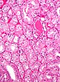
Microscopic
Classic
Features - classic type (3 P's mnemonic):[3][2]
- Pale cytoplasm, with wisps of eosinophilic material; the cells are not completely clear, they have "cobwebs".
- Perinuclear clearing, i.e. a pale halo surrounds the nucleus - key feature.
- Periphery of cell distinct, i.e. cell membrane is easy to discern.
Notes:
- May have psammoma bodies.
- May be described as "plant-like"; plant cells have (thick) cell walls.
- The perinuclear clearing is often somewhat patchy, i.e. it is usually not present in very tumour cell.
DDx:
- Clear cell RCC (classic).
- Perinuclear clearing is not seen in clear cell RCC.
- ChRCC has wisps in the cytoplasm.
Eosinophilic variant
Features - eosinophilic variant:[2]
- Eosinophilic (finely granular) cytoplasm.
- Perinuclear clearing - key feature.
- Periphery of cell distinct.
- Smaller cells than classic subtype.
Notes:
- May have psammoma bodies.
DDx:
- Oncocytoma - particularly the eosinophilic variant.
- IHC may be useful to differentiate (CK7: oncocytoma = cytoplasm +ve, chromophobe = cell membrane +ve).
- A comparison based on histomorphology: Tabular comparison between ChRCC & oncocytoma.
- Oncocytoma typically has: no perinuclear clearing, no raisinoid nuclei, no binucleation.
- Clear cell RCC, eosinophilic variant.
- Perinuclear clearing is not seen in clear cell RCC.
- ChRCC has wisps in the cytoplasm.
Image
Oncocytic chromophobe RCC. (WC/Nephron)

www:
Stains
- Hale's colloidal iron +ve (blue granular cytoplasmic).
Images:
- ChRCC Hale's colloidal iron - several images (nature.com).
- ChRCC Hale's colloidal iron (ultrapath.org).[4]
- ChRCC Hale's colloidal iron (diagnosticpathology.org).
IHC
- CK7 +ve cell membrane.[2]
- CD117 +ve.
- Vimentin -ve.
Molecular
- Extensive aneusomy (monosomy?):[5]
- Loss of chromosomes: 1, 2, 6, 10, 13, 17, 21.
Sign out
KIDNEY, RIGHT UPPER POLE, PARTIAL NEPHRECTOMY:
- CHROMOPHOBE RENAL CELL CARCINOMA.

COMMENT:
The sections show a mix of clear cells with wispy cytoplasm, and cells with eosinophilic
cytoplasm and perinuclear halos. There are no true papillae.

Stains and immunostains:
Positive: CK7, CAM5.2, EMA, pankeratin, CD117, colloidal iron.
Negative: AMACR, CD10, CD68, RCC, vimentin.
See also
References
- ↑ Delahunt, B.; Sika-Paotonu, D.; Bethwaite, PB.; McCredie, MR.; Martignoni, G.; Eble, JN.; Jordan, TW. (Jun 2007). "Fuhrman grading is not appropriate for chromophobe renal cell carcinoma.". Am J Surg Pathol 31 (6): 957-60. doi:10.1097/01.pas.0000249446.28713.53. PMID 17527087.
- ↑ 2.0 2.1 2.2 2.3 Zhou, Ming; Magi-Galluzzi, Cristina (2006). Genitourinary Pathology: A Volume in Foundations in Diagnostic Pathology Series (1st ed.). Churchill Livingstone. pp. 293. ISBN 978-0443066771.
- ↑ Cotran, Ramzi S.; Kumar, Vinay; Fausto, Nelson; Nelso Fausto; Robbins, Stanley L.; Abbas, Abul K. (2005). Robbins and Cotran pathologic basis of disease (7th ed.). St. Louis, Mo: Elsevier Saunders. pp. 1016-7. ISBN 0-7216-0187-1.
- ↑ URL: http://www.ultrapath.org/oldsite/cases99/sep99/cotm9-2.html. Accessed on: 9 October 2011.
- ↑ Humphrey, Peter A; Dehner, Louis P; Pfeifer, John D (2008). The Washington Manual of Surgical Pathology (1st ed.). Lippincott Williams & Wilkins. pp. 292. ISBN 978-0781765275.