Difference between revisions of "Chordoma"
Jump to navigation
Jump to search
| Line 57: | Line 57: | ||
*[[Myxoid lesions]]. | *[[Myxoid lesions]]. | ||
**Myxopapillary ependymoma. | **Myxopapillary ependymoma. | ||
**Myxoid liposarcoma - negative for EMA and cytokeratins. | **[[Myxoid liposarcoma]] - negative for EMA and cytokeratins. | ||
* | *Choroid lesions: | ||
**Choroid meningioma. | **[[Choroid meningioma]]. | ||
**Choroid glioma - location, location, location. | **Choroid glioma - location, location, location. | ||
**Large notochordal rest - only evidence of destructive growth can identify a chordoma. | **Large notochordal rest - only evidence of destructive growth can identify a chordoma. | ||
*Metastasis | *[[Metastasis]]: | ||
**Metastatic signet ring cell | **Metastatic [[signet ring cell carcinoma]] - negative for S100 and brachyury; clinical history (important). | ||
**Metastatic clear cell renal cell carcinoma - negative for S100 and brachyury ( | **Metastatic [[clear cell renal cell carcinoma]] - negative for S100 and brachyury; clinical history (important). | ||
*[[Parachordoma]] - extremely rare. | *[[Parachordoma]] - extremely rare. | ||
Revision as of 17:12, 4 December 2014
| Chordoma | |
|---|---|
| Diagnosis in short | |
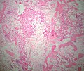
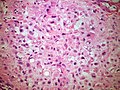
 Chordoma. HPS stain. | |
|
| |
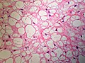
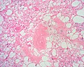
| LM | physaliphorous cells (also bubble cells) - very large clear bubble with a sharp border, bubble does not compress nucleus; islands of cells surrounded by fibrous tissue; myxoid background |
| LM DDx | chondrosarcoma, myxoid lesions, parachordoma |
| IHC | S-100 +ve, AE1/AE3 +ve, Brachyury +ve, EMA +ve |
| Gross | myxoid |
| Site | sacrum or clivus |
|
| |
| Prevalence | uncommon |
Chordoma is an uncommon tumour in neuropathology.
General
- Location: usually sacrum or clivus.
- It is a bone tumour.
Gross
- Soft, gelatinous, lobulated.[1]
DDx:
- Bony metastasis (mucinous carcinoma) - typically multifocal.
Image:
Microscopic
Features:[2]
- Architecture: islands of cells surrounded by fibrous tissue.
- Also described as "lobulated" architecture; may not be apparent.
- Myxoid background - grey extracellular material, variable amount present.
- Mixed cell population:
- Abundant eosinophilic cytoplasm.
- Physaliphorous cells or bubble cells - key feature.
- Have a very large clear bubble with a sharp border; bubble does not compress nucleus - nucleus may be in bubble.
DDx:
- Chondrosarcoma - negative for EMA and cytokeratins. Beware of 'chondroid' chordoma.
- Myxoid lesions.
- Myxopapillary ependymoma.
- Myxoid liposarcoma - negative for EMA and cytokeratins.
- Choroid lesions:
- Choroid meningioma.
- Choroid glioma - location, location, location.
- Large notochordal rest - only evidence of destructive growth can identify a chordoma.
- Metastasis:
- Metastatic signet ring cell carcinoma - negative for S100 and brachyury; clinical history (important).
- Metastatic clear cell renal cell carcinoma - negative for S100 and brachyury; clinical history (important).
- Parachordoma - extremely rare.
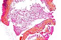
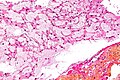
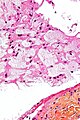
Images

Chordoma - low mag. (WC)
Chordoma - intermed. mag. (WC)
Chordoma - high mag. (WC)
Chordoma - very high mag. (WC)
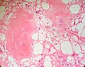
Physaliphorous cells. (SKB)
Low power view. Somewhat lobulated tumour with loose areas, cellular areas and fibrous septa. (SKB)
Physaliphorous cells. (SKB)
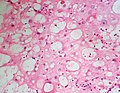
Physaliphorous cells. (SKB)
Physaliphorous cells.(SKB)
Chordoma cells may form sheets of cells with eosinophilic cytoplasm. (SKB)


www:
- Chordoma (upmc.edu).
- Chordoma - sacrum - several images (upmc.edu).
- Chordoma - additional case with several images (upmc.edu).
IHC
- S-100 +ve.
- AE1/AE3 +ve.
- Brachyury +ve -- key stain.
- EMA +ve.
Key points:
- Brachyury is not a commonly stocked antibody.
- Chordoma will be S100 AND Epithelial marker positive.
- Many other items in the DDX will be either S100 OR Epithelial marker positive.
See also
References
- ↑ URL: http://www.histopathology-india.net/Chordoma.htm. Accessed on: 12 April 2012.
- ↑ Tadrous, Paul.J. Diagnostic Criteria Handbook in Histopathology: A Surgical Pathology Vade Mecum (1st ed.). Wiley. pp. 184. ISBN 978-0470519035.
- ↑ URL: http://path.upmc.edu/cases/case312/micro.html. Accessed on: 14 January 2012.
- ↑ Coindre, JM.; Rivel, J.; Trojani, M.; De Mascarel, I.; De Mascarel, A. (Sep 1986). "Immunohistological study in chordomas.". J Pathol 150 (1): 61-3. doi:10.1002/path.1711500110. PMID 2431128.
- ↑ Online 'Mendelian Inheritance in Man' (OMIM) 601397
- ↑ URL: http://www.jstor.org/pss/86845. Accessed on: 18 May 2010.