Difference between revisions of "Cardiac tumours"
m (→General) |
|||
| (45 intermediate revisions by 2 users not shown) | |||
| Line 1: | Line 1: | ||
'''Cardiac tumours''' are rare buggers. | '''[[Heart|Cardiac]] tumours''' are rare buggers. They provide some work for cardiac surgeons. | ||
=Most common malignant= | |||
*Metastases - most common cardiac tumour; 1:~100 primary tumours:secondary tumours.<ref name=pmid20472615>{{Cite journal | last1 = Castillo | first1 = JG. | last2 = Silvay | first2 = G. | title = Characterization and management of cardiac tumors. | journal = Semin Cardiothorac Vasc Anesth | volume = 14 | issue = 1 | pages = 6-20 | month = Mar | year = 2010 | doi = 10.1177/1089253210362596 | PMID = 20472615 | *[[Metastases]] - most common cardiac tumour; 1:~100 primary tumours:secondary tumours.<ref name=pmid20472615>{{Cite journal | last1 = Castillo | first1 = JG. | last2 = Silvay | first2 = G. | title = Characterization and management of cardiac tumors. | journal = Semin Cardiothorac Vasc Anesth | volume = 14 | issue = 1 | pages = 6-20 | month = Mar | year = 2010 | doi = 10.1177/1089253210362596 | PMID = 20472615 }}</ref> | ||
=Primary heart tumours= | |||
*Approximately 10% of resected (primary) cardiac tumours are malignant.<ref name=pmid18350921>{{cite journal |author=Burke A |title=Primary malignant cardiac tumors |journal=Semin Diagn Pathol |volume=25 |issue=1 |pages=39-46 |year=2008 |month=February |pmid=18350921 |doi= |url=}}</ref> | *Approximately 10% of resected (primary) cardiac tumours are malignant.<ref name=pmid18350921>{{cite journal |author=Burke A |title=Primary malignant cardiac tumors |journal=Semin Diagn Pathol |volume=25 |issue=1 |pages=39-46 |year=2008 |month=February |pmid=18350921 |doi= |url=}}</ref> | ||
**90% are sarcomas. | **90% are sarcomas. | ||
| Line 11: | Line 10: | ||
In order of frequency: | In order of frequency: | ||
#Myxoma. | #[[Cardiac myxoma|Myxoma]]. | ||
#[[Lipoma]]. | #[[Lipoma]]. | ||
#Papillary fibroelastoma. | #[[Papillary fibroelastoma]]. | ||
Notes: | Notes: | ||
| Line 20: | Line 19: | ||
Malignant heart tumours (in order of frequency):<ref name=pmid20472615>{{Cite journal | last1 = Castillo | first1 = JG. | last2 = Silvay | first2 = G. | title = Characterization and management of cardiac tumors. | journal = Semin Cardiothorac Vasc Anesth | volume = 14 | issue = 1 | pages = 6-20 | month = Mar | year = 2010 | doi = 10.1177/1089253210362596 | PMID = 20472615 }}</ref> | Malignant heart tumours (in order of frequency):<ref name=pmid20472615>{{Cite journal | last1 = Castillo | first1 = JG. | last2 = Silvay | first2 = G. | title = Characterization and management of cardiac tumors. | journal = Semin Cardiothorac Vasc Anesth | volume = 14 | issue = 1 | pages = 6-20 | month = Mar | year = 2010 | doi = 10.1177/1089253210362596 | PMID = 20472615 }}</ref> | ||
#Undifferentiated. | #Undifferentiated. | ||
#Angiosarcoma. | #[[Angiosarcoma]]. | ||
#Leiomyosarcoma. | #[[Leiomyosarcoma]]. | ||
Note: | |||
*According to WMSP:<ref name=Ref_WMSP135>{{Ref WMSP|135}}</ref> Most common primary malignant tumour of the heart = angiosarcoma. | |||
* | |||
=Specific entities= | |||
==Cardiac myxoma== | |||
{{Main|Cardiac myxoma}} | |||
This includes the common ''atrial myxoma''. | |||
== | ==Papillary fibroelastoma== | ||
*[[AKA]] ''fibroelastoma''. | |||
**Should '''not''' be confused with ''[[elastofibroma]]''. | |||
*[[AKA]] ''giant Lambl excrescence''.<ref name=pmid9767897>{{Cite journal | last1 = Loire | first1 = R. | last2 = Pinède | first2 = L. | last3 = Donsbeck | first3 = AV. | last4 = Nighoghossian | first4 = N. | last5 = Perinetti | first5 = M. | title = [Papillary fibroelastoma of the heart (giant Lambl excrescence). Clinical-anatomical study on 10 surgically treated patients]. | journal = Presse Med | volume = 27 | issue = 16 | pages = 753-7 | month = Apr | year = 1998 | doi = | PMID = 9767897 }}</ref> | |||
===General=== | |||
*Usually an incidental finding. | |||
* | |||
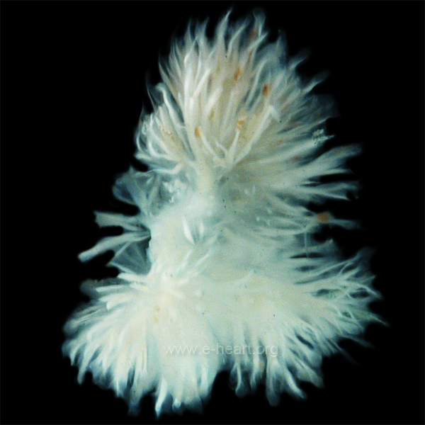
=== | ===Gross=== | ||
*Friable appearing. | |||
*Yellow. | |||
* | *Typically on free edge. | ||
* | |||
* | |||
Image: | |||
*[http:// | *[http://www.e-heart.org/Photos/09_Cardiac_Tumors_Photos/%C2%A9%20Papillary%20fibroelastoma%20-%20Gross.jpg Papillary fibroelastoma (e-heart.org)].<ref>URL: [http://www.e-heart.org/pages/09_Cardiac_Tumors/09_Cardiac_Tumors_Primary_Benign_Cardiac_Papillary%20Fibroelastoma_001.htm http://www.e-heart.org/pages/09_Cardiac_Tumors/09_Cardiac_Tumors_Primary_Benign_Cardiac_Papillary%20Fibroelastoma_001.htm]. 5 March 2013.</ref> | ||
===Microscopic=== | ===Microscopic=== | ||
Features:<ref>[http://www.pathologyoutlines.com/hearttumor.html http://www.pathologyoutlines.com/hearttumor.html]</ref> | Features:<ref>URL: [http://www.pathologyoutlines.com/hearttumor.html http://www.pathologyoutlines.com/hearttumor.html]. Accessed on: 16 May 2011.</ref> | ||
*Braching papillary fronds which are: | *Braching papillary fronds which are: | ||
**Composed of collagen, and | **Composed of collagen, and | ||
| Line 72: | Line 56: | ||
**Mucopolysaccharide. | **Mucopolysaccharide. | ||
====Images==== | |||
<gallery> | |||
Image:Papillary_fibroelastoma.jpg | Papillary fibroelastoma - low mag. (WC) | |||
Image:Papillary_fibroelastoma2.jpg | Papillary fibroelastoma - intermed. mag. (WC) | |||
</gallery> | |||
== | ==Cardiac rhabdomyoma== | ||
{{Main|Rhabdomyoma}} | |||
*Very rare. | *Very rare. | ||
*Benign. | *Benign. | ||
| Line 82: | Line 69: | ||
==Rhabdomyosarcoma== | ==Rhabdomyosarcoma== | ||
See ''[[ | See ''[[rhabdomyosarcoma]]''. | ||
==Lipoma== | ==Lipoma== | ||
| Line 92: | Line 79: | ||
*Lipomatous hypertrophy.<ref name=pmid20196664>{{Cite journal | last1 = Miller | first1 = DV. | last2 = Tazelaar | first2 = HD. | title = Cardiovascular pseudoneoplasms. | journal = Arch Pathol Lab Med | volume = 134 | issue = 3 | pages = 362-8 | month = Mar | year = 2010 | doi = | PMID = 20196664 }}</ref> | *Lipomatous hypertrophy.<ref name=pmid20196664>{{Cite journal | last1 = Miller | first1 = DV. | last2 = Tazelaar | first2 = HD. | title = Cardiovascular pseudoneoplasms. | journal = Arch Pathol Lab Med | volume = 134 | issue = 3 | pages = 362-8 | month = Mar | year = 2010 | doi = | PMID = 20196664 }}</ref> | ||
==Cystic | ==Cystic tumour of the atrioventricular nodal region== | ||
{{Main|Cystic tumour of the atrioventricular nodal region}} | |||
== | |||
== | ==Angiosarcoma== | ||
[[File: Angiosarc heart 1 sl 1.png| Angiosarcoma of right atrium]] | |||
[[File: Angiosarc heart 1 sl 2.png| Angiosarcoma of right atrium]] | |||
[[File: Angiosarc heart 1 sl 3.png| Angiosarcoma of right atrium]] | |||
[[File: Angiosarc heart 1 sl 4.png| Angiosarcoma of right atrium]] | |||
[[File: Angiosarc heart 1 sl 5.png| Angiosarcoma of right atrium]] | |||
[[File: Angiosarc heart 1 sl 6.png| Angiosarcoma of right atrium]] | |||
Angiosarcoma of right atrium. A. Luminal clot tops tumor with blood filled spaces. B. Tumor replaces myocardium. C. Irregularly branched vessels, sometimes associated with extravasated erythrocytes. D. Irregularly aggregated and oriented spindle cells. E. Cells vary in size and shape much more so than an organizing thrombus or hemangioma. Note luminal tufts (green arrows), clustered large cancer nuclei with nucleoli (blue arrows). F. Cytoplasmic vacuoles, some with erythrocytes (arrows). | |||
=See also= | |||
*[[Heart]]. | *[[Heart]]. | ||
*[[Valvular heart disease]]. | *[[Valvular heart disease]]. | ||
=References= | |||
{{reflist|2}} | {{reflist|2}} | ||
[[Category:Cardiovascular pathology]] | [[Category:Cardiovascular pathology]] | ||
[[Category:Cardiac tumours]] | |||
Latest revision as of 19:04, 24 March 2019
Cardiac tumours are rare buggers. They provide some work for cardiac surgeons.
Most common malignant
- Metastases - most common cardiac tumour; 1:~100 primary tumours:secondary tumours.[1]
Primary heart tumours
- Approximately 10% of resected (primary) cardiac tumours are malignant.[2]
- 90% are sarcomas.
- 10% are lymphomas.
In order of frequency:
Notes:
- If one is considering only valves - papillary fibroelastoma is No. 1.
Malignant heart tumours (in order of frequency):[1]
- Undifferentiated.
- Angiosarcoma.
- Leiomyosarcoma.
Note:
- According to WMSP:[3] Most common primary malignant tumour of the heart = angiosarcoma.
Specific entities
Cardiac myxoma
This includes the common atrial myxoma.
Papillary fibroelastoma
- AKA fibroelastoma.
- Should not be confused with elastofibroma.
- AKA giant Lambl excrescence.[4]
General
- Usually an incidental finding.
Gross
- Friable appearing.
- Yellow.
- Typically on free edge.
Image:
Microscopic
Features:[6]
- Braching papillary fronds which are:
- Composed of collagen, and
- Avascular.
- +/-Elastic tissue.
- Surrounded by:
- Endothelium, and
- Mucopolysaccharide.
Images
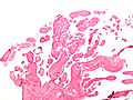
Papillary fibroelastoma - low mag. (WC)
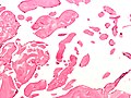
Papillary fibroelastoma - intermed. mag. (WC)


Cardiac rhabdomyoma
- Very rare.
- Benign.
- Associated with tuberous sclerosis.[7]
Rhabdomyosarcoma
See rhabdomyosarcoma.
Lipoma
- Like lipomas elsewhere in the body.
- Usually location: left ventricle, subendocardial.[3]
DDx:
- Lipomatous hypertrophy.[8]
Cystic tumour of the atrioventricular nodal region
Angiosarcoma


Angiosarcoma of right atrium. A. Luminal clot tops tumor with blood filled spaces. B. Tumor replaces myocardium. C. Irregularly branched vessels, sometimes associated with extravasated erythrocytes. D. Irregularly aggregated and oriented spindle cells. E. Cells vary in size and shape much more so than an organizing thrombus or hemangioma. Note luminal tufts (green arrows), clustered large cancer nuclei with nucleoli (blue arrows). F. Cytoplasmic vacuoles, some with erythrocytes (arrows).
See also
References
- ↑ 1.0 1.1 Castillo, JG.; Silvay, G. (Mar 2010). "Characterization and management of cardiac tumors.". Semin Cardiothorac Vasc Anesth 14 (1): 6-20. doi:10.1177/1089253210362596. PMID 20472615.
- ↑ Burke A (February 2008). "Primary malignant cardiac tumors". Semin Diagn Pathol 25 (1): 39-46. PMID 18350921.
- ↑ 3.0 3.1 Humphrey, Peter A; Dehner, Louis P; Pfeifer, John D (2008). The Washington Manual of Surgical Pathology (1st ed.). Lippincott Williams & Wilkins. pp. 135. ISBN 978-0781765275.
- ↑ Loire, R.; Pinède, L.; Donsbeck, AV.; Nighoghossian, N.; Perinetti, M. (Apr 1998). "[Papillary fibroelastoma of the heart (giant Lambl excrescence). Clinical-anatomical study on 10 surgically treated patients].". Presse Med 27 (16): 753-7. PMID 9767897.
- ↑ URL: http://www.e-heart.org/pages/09_Cardiac_Tumors/09_Cardiac_Tumors_Primary_Benign_Cardiac_Papillary%20Fibroelastoma_001.htm. 5 March 2013.
- ↑ URL: http://www.pathologyoutlines.com/hearttumor.html. Accessed on: 16 May 2011.
- ↑ Yinon, Y.; Chitayat, D.; Blaser, S.; Seed, M.; Amsalem, H.; Yoo, SJ.; Jaeggi, ET. (Aug 2010). "Fetal cardiac tumors: a single-center experience of 40 cases.". Prenat Diagn. doi:10.1002/pd.2590. PMID 20721876.
- ↑ Miller, DV.; Tazelaar, HD. (Mar 2010). "Cardiovascular pseudoneoplasms.". Arch Pathol Lab Med 134 (3): 362-8. PMID 20196664.