Difference between revisions of "Cardiac tumours"
Jump to navigation
Jump to search
(→Cardiac myxoma: tweak) |
|||
| Line 27: | Line 27: | ||
=Specific entities= | =Specific entities= | ||
==Cardiac myxoma== | ==Cardiac myxoma== | ||
{{Main|Cardiac myxoma}} | |||
This includes the common ''atrial myxoma''. | |||
==Papillary fibroelastoma== | ==Papillary fibroelastoma== | ||
Revision as of 06:40, 3 October 2013
Cardiac tumours are rare buggers. They provide some work for cardiac surgeons.
Most common malignant
- Metastases - most common cardiac tumour; 1:~100 primary tumours:secondary tumours.[1]
Primary heart tumours
- Approximately 10% of resected (primary) cardiac tumours are malignant.[2]
- 90% are sarcomas.
- 10% are lymphomas.
In order of frequency:
Notes:
- If one is considering only valves - papillary fibroelastoma is No. 1.
Malignant heart tumours (in order of frequency):[1]
- Undifferentiated.
- Angiosarcoma.
- Leiomyosarcoma.
Note:
- According to WMSP:[3] Most common primary malignant tumour of the heart = angiosarcoma.
Specific entities
Cardiac myxoma
Main article: Cardiac myxoma
This includes the common atrial myxoma.
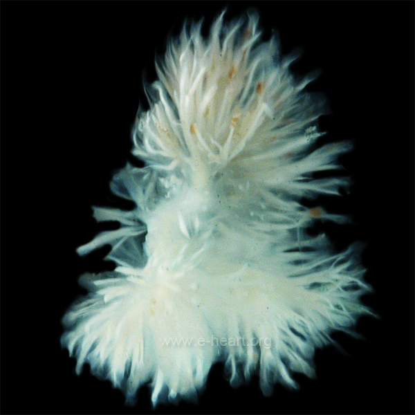
Papillary fibroelastoma
General
- Usually an incidental finding.
Gross
- Friable appearing.
- Yellow.
- Typically on free edge.
Image:
Microscopic
Features:[6]
- Braching papillary fronds which are:
- Composed of collagen, and
- Avascular.
- +/-Elastic tissue.
- Surrounded by:
- Endothelium, and
- Mucopolysaccharide.
Images
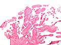
Papillary fibroelastoma - low mag. (WC)
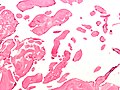
Papillary fibroelastoma - intermed. mag. (WC)


Cardiac rhabdomyoma
Main article: Rhabdomyoma
- Very rare.
- Benign.
- Associated with tuberous sclerosis.[7]
Rhabdomyosarcoma
See soft tissue tumours.
Lipoma
Main article: Adipocytic tumours
- Like lipomas elsewhere in the body.
- Usually location: left ventricle, subendocardial.[3]
DDx:
- Lipomatous hypertrophy.[8]
Cystic tumour of the atrioventricular nodal region
General
- Super rare.[9]
- Usually 1-2 mm.
- May cause sudden cardiac death.[10]
Gross
- "Bump" in the triangle of Koch.
- Cystic spaces.
Microscopic
Features:[10]
- Cystic spaces lined single layer of epithelial cells.
- +/-Focal mononuclear inflammation.
- +/-Psammoma bodies.
Images
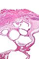
CTAVNR - very low mag. (WC)
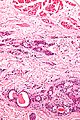
CTAVNR - high mag. (WC)


IHC
- CEA +ve.
- EMA +ve.
See also
References
- ↑ 1.0 1.1 Castillo, JG.; Silvay, G. (Mar 2010). "Characterization and management of cardiac tumors.". Semin Cardiothorac Vasc Anesth 14 (1): 6-20. doi:10.1177/1089253210362596. PMID 20472615.
- ↑ Burke A (February 2008). "Primary malignant cardiac tumors". Semin Diagn Pathol 25 (1): 39-46. PMID 18350921.
- ↑ 3.0 3.1 Humphrey, Peter A; Dehner, Louis P; Pfeifer, John D (2008). The Washington Manual of Surgical Pathology (1st ed.). Lippincott Williams & Wilkins. pp. 135. ISBN 978-0781765275.
- ↑ Loire, R.; Pinède, L.; Donsbeck, AV.; Nighoghossian, N.; Perinetti, M. (Apr 1998). "[Papillary fibroelastoma of the heart (giant Lambl excrescence). Clinical-anatomical study on 10 surgically treated patients].". Presse Med 27 (16): 753-7. PMID 9767897.
- ↑ URL: http://www.e-heart.org/pages/09_Cardiac_Tumors/09_Cardiac_Tumors_Primary_Benign_Cardiac_Papillary%20Fibroelastoma_001.htm. 5 March 2013.
- ↑ URL: http://www.pathologyoutlines.com/hearttumor.html. Accessed on: 16 May 2011.
- ↑ Yinon, Y.; Chitayat, D.; Blaser, S.; Seed, M.; Amsalem, H.; Yoo, SJ.; Jaeggi, ET. (Aug 2010). "Fetal cardiac tumors: a single-center experience of 40 cases.". Prenat Diagn. doi:10.1002/pd.2590. PMID 20721876.
- ↑ Miller, DV.; Tazelaar, HD. (Mar 2010). "Cardiovascular pseudoneoplasms.". Arch Pathol Lab Med 134 (3): 362-8. PMID 20196664.
- ↑ Kaminishi, Y.; Watanabe, Y.; Nakata, H.; Shimokama, T.; Jikuya, T. (Jan 2002). "Cystic tumor of the atrioventricular nodal region.". Jpn J Thorac Cardiovasc Surg 50 (1): 37-9. PMID 11855098.
- ↑ 10.0 10.1 Paniagua, JR.; Sadaba, JR.; Davidson, LA.; Munsch, CM. (Apr 2000). "Cystic tumour of the atrioventricular nodal region: report of a case successfully treated with surgery.". Heart 83 (4): E6. PMID 10722558.