Difference between revisions of "Cardiac tumours"
(→Cardiac myxoma: tweak) |
|||
| Line 67: | Line 67: | ||
*[[Myxoid liposarcoma]] - chickenwire vasculature. | *[[Myxoid liposarcoma]] - chickenwire vasculature. | ||
Images | ====Images==== | ||
<gallery> | |||
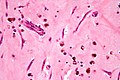
Image:Atrial_myxoma_high_mag.jpg | Atrial myxoma - high mag. (WC) | |||
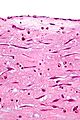
Image:Atrial_myxoma_edge_high_mag.jpg | Atrial myxoma - endothelial covering - high mag. (WC) | |||
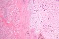
Image:Atrial_myxoma_edge_low_mag.jpg | Atrial myxoma - sharp border - low mag. (WC) | |||
</gallery> | |||
www: | |||
*[http://radiographics.rsna.org/content/22/3/673/F3.expansion.html Atrial myxoma (rsna.org)].<ref name=pmid12006696/> | |||
*[http://radiographics.rsna.org/content/22/3/673/F4.expansion.html Gamna bodies (rsna.org)].<ref name=pmid12006696/> | |||
===Sign out=== | ===Sign out=== | ||
Revision as of 06:28, 3 October 2013
Cardiac tumours are rare buggers. They provide some work for cardiac surgeons.
Most common malignant
- Metastases - most common cardiac tumour; 1:~100 primary tumours:secondary tumours.[1]
Primary heart tumours
- Approximately 10% of resected (primary) cardiac tumours are malignant.[2]
- 90% are sarcomas.
- 10% are lymphomas.
In order of frequency:
Notes:
- If one is considering only valves - papillary fibroelastoma is No. 1.
Malignant heart tumours (in order of frequency):[1]
- Undifferentiated.
- Angiosarcoma.
- Leiomyosarcoma.
Note:
- According to WMSP:[3] Most common primary malignant tumour of the heart = angiosarcoma.
Specific entities
Cardiac myxoma
General
- Uncommon.
- Clinical: may lead to cerebral infarction.[4]
- Diagnosed by imaging.
- May be familial, i.e. Carney complex (AKA NAME syndrome, AKA LAMB syndrome).[5]
- NAME = Nevi, Atrial myxoma, Myxoid neurofibroma, and Ephelides (freckles[6]).
- LAMB = Lentigines, Atrial myxomas, Mucocutaneous myxomas, Blue nevi.
Most common presentations:[4]
- Dyspnea - 45%.
- Neurologic symptoms 36%.
Gross
Location:[4]
- Usually atrial.
- Usually left side ~60%.[7]
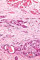
Features:[7]
- Lobular surface.
- Smooth surface.
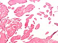
Microscopic
Features:[7]
- Myxoid material - extra cellular - key feature.
- Myxoma cells:[8]
- Stellate, polygonal or spindled morphology.
- +/-Multinucleated.
- Inconspicuous nucleoli.
- Abundant cytoplasm.
- Calcified elastic fibers - gamna bodies.
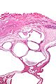
- Hemorrhage common.
- Often covered by endothelium.
- +/-Ossification.
- +/-Fibrosis.
DDx:[9]
- Papillary fibroelastoma - not really in the histomorphologic differential diagnosis.
- Myxofibrosarcoma - nuclear pleomorphism.
- Myxoid liposarcoma - chickenwire vasculature.
Images
Atrial myxoma - high mag. (WC)
Atrial myxoma - endothelial covering - high mag. (WC)
Atrial myxoma - sharp border - low mag. (WC)


www:
Sign out
MASS, LEFT ATRIUM, EXCISION:
- MYXOMA.
Micro
The sections show paucicellular myxoid material containing polygonal and spindled cells with eosinophilic myxoid cytoplasm, bland nuclei, inconspicuous nucleoli and focal multinucleation (myxoma cells). Hemosiderin-laden macrophages, calcified elastic fibres (gamna bodies) and scattered inflammatory cells are also present. There is no nuclear atypia. Mitotic activity is not evident. Several sections show fresh hemorrhage. The edge has a fibrotic rim and appears to be covered by endothelium. No cardiac muscle is identified.
Papillary fibroelastoma
General
- Usually an incidental finding.
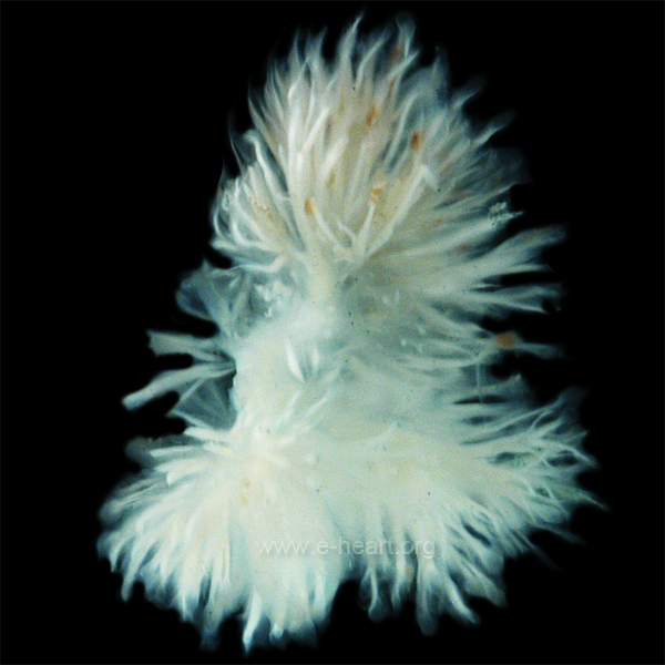
Gross
- Friable appearing.
- Yellow.
- Typically on free edge.
Image:
Microscopic
Features:[12]
- Braching papillary fronds which are:
- Composed of collagen, and
- Avascular.
- +/-Elastic tissue.
- Surrounded by:
- Endothelium, and
- Mucopolysaccharide.
Images
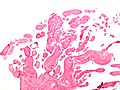
Papillary fibroelastoma - low mag. (WC)
Papillary fibroelastoma - intermed. mag. (WC)


Cardiac rhabdomyoma
- Very rare.
- Benign.
- Associated with tuberous sclerosis.[13]
Rhabdomyosarcoma
See soft tissue tumours.
Lipoma
- Like lipomas elsewhere in the body.
- Usually location: left ventricle, subendocardial.[3]
DDx:
- Lipomatous hypertrophy.[14]
Cystic tumour of the atrioventricular nodal region
General
- Super rare.[15]
- Usually 1-2 mm.
- May cause sudden cardiac death.[16]
Gross
- "Bump" in the triangle of Koch.
- Cystic spaces.
Microscopic
Features:[16]
- Cystic spaces lined single layer of epithelial cells.
- +/-Focal mononuclear inflammation.
- +/-Psammoma bodies.
Images
CTAVNR - very low mag. (WC)
CTAVNR - high mag. (WC)


IHC
- CEA +ve.
- EMA +ve.
See also
References
- ↑ 1.0 1.1 Castillo, JG.; Silvay, G. (Mar 2010). "Characterization and management of cardiac tumors.". Semin Cardiothorac Vasc Anesth 14 (1): 6-20. doi:10.1177/1089253210362596. PMID 20472615.
- ↑ Burke A (February 2008). "Primary malignant cardiac tumors". Semin Diagn Pathol 25 (1): 39-46. PMID 18350921.
- ↑ 3.0 3.1 Humphrey, Peter A; Dehner, Louis P; Pfeifer, John D (2008). The Washington Manual of Surgical Pathology (1st ed.). Lippincott Williams & Wilkins. pp. 135. ISBN 978-0781765275.
- ↑ 4.0 4.1 4.2 Knepper LE, Biller J, Adams HP, Bruno A (November 1988). "Neurologic manifestations of atrial myxoma. A 12-year experience and review". Stroke 19 (11): 1435-40. PMID 3188128. http://stroke.ahajournals.org/cgi/reprint/19/11/1435.
- ↑ Humphrey, Peter A; Dehner, Louis P; Pfeifer, John D (2008). The Washington Manual of Surgical Pathology (1st ed.). Lippincott Williams & Wilkins. pp. 135. ISBN 978-0781765275.
- ↑ URL: http://emedicine.medscape.com/article/1119293-overview. Accessed on: 7 January 2011.
- ↑ 7.0 7.1 7.2 7.3 7.4 Grebenc, ML.; Rosado-de-Christenson, ML.; Green, CE.; Burke, AP.; Galvin, JR.. "Cardiac myxoma: imaging features in 83 patients.". Radiographics 22 (3): 673-89. PMID 12006696. Cite error: Invalid
<ref>tag; name "pmid12006696" defined multiple times with different content - ↑ Orlandi, A.; Ciucci, A.; Ferlosio, A.; Genta, R.; Spagnoli, LG.; Gabbiani, G. (Jun 2006). "Cardiac myxoma cells exhibit embryonic endocardial stem cell features.". J Pathol 209 (2): 231-9. doi:10.1002/path.1959. PMID 16508920.
- ↑ Tadrous, Paul.J. Diagnostic Criteria Handbook in Histopathology: A Surgical Pathology Vade Mecum (1st ed.). Wiley. pp. 79. ISBN 978-0470519035.
- ↑ Loire, R.; Pinède, L.; Donsbeck, AV.; Nighoghossian, N.; Perinetti, M. (Apr 1998). "[Papillary fibroelastoma of the heart (giant Lambl excrescence). Clinical-anatomical study on 10 surgically treated patients].". Presse Med 27 (16): 753-7. PMID 9767897.
- ↑ URL: http://www.e-heart.org/pages/09_Cardiac_Tumors/09_Cardiac_Tumors_Primary_Benign_Cardiac_Papillary%20Fibroelastoma_001.htm. 5 March 2013.
- ↑ URL: http://www.pathologyoutlines.com/hearttumor.html. Accessed on: 16 May 2011.
- ↑ Yinon, Y.; Chitayat, D.; Blaser, S.; Seed, M.; Amsalem, H.; Yoo, SJ.; Jaeggi, ET. (Aug 2010). "Fetal cardiac tumors: a single-center experience of 40 cases.". Prenat Diagn. doi:10.1002/pd.2590. PMID 20721876.
- ↑ Miller, DV.; Tazelaar, HD. (Mar 2010). "Cardiovascular pseudoneoplasms.". Arch Pathol Lab Med 134 (3): 362-8. PMID 20196664.
- ↑ Kaminishi, Y.; Watanabe, Y.; Nakata, H.; Shimokama, T.; Jikuya, T. (Jan 2002). "Cystic tumor of the atrioventricular nodal region.". Jpn J Thorac Cardiovasc Surg 50 (1): 37-9. PMID 11855098.
- ↑ 16.0 16.1 Paniagua, JR.; Sadaba, JR.; Davidson, LA.; Munsch, CM. (Apr 2000). "Cystic tumour of the atrioventricular nodal region: report of a case successfully treated with surgery.". Heart 83 (4): E6. PMID 10722558.