Canalicular adenoma
Jump to navigation
Jump to search
| Canalicular adenoma | |
|---|---|
| Diagnosis in short | |
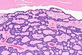
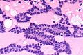
 Canalicular adenoma. H&E stain. (WC) | |
|
| |
| LM |
cords of tumour ("canals") with beading (characteristic), cystic spaces/tubules, intraluminal squamous balls (common) |
| Site | salivary gland - usually upper lip or buccal mucosa |
|
| |
| Signs | mass lesion |
| Prevalence | very rare |
Canalicular adenoma is a rare salivary gland tumour.
General
- Exclusively oral cavity.
Clinical:
- Mass lesion.[1]
Gross
- Classically upper lip - may be buccal mucosa or palate.
Note:
- In one large series of 67 cases:[1]
- Upper lip 69% (47/67).
- Buccal mucosa 25% (17/67).
- Palate 6% (4/67).
Microscopic
Features:[1]
- Cords of tumour ("canals") with beading - characteristic.
- Cystic spaces/tubules.
- Intraluminal squamous balls - common (~60% of cases).
- Myxoid/bluish stroma.
DDx:
Images
CA - intermed. mag. (WC)
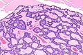
CA - intermed. mag. (WC)
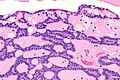
CA - high mag. (WC)
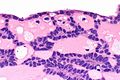
CA - very high mag. (WC)
CA - very high mag. (WC)


www
IHC
Features:[1]
- S-100 +ve - diffuse and strong.
- Pankeratin +ve - diffuse and strong.
- GFAP +ve - luminal.
- SOX10 +ve - nuclear.
- p16 +ve - luminal squamous balls.
- CK5/6 +ve - luminal squamous balls.
- p63 -ve.
- Nuclei negative, cytoplasm positive.
- Positive in basal cell adenoma.
See also
References
- ↑ 1.0 1.1 1.2 1.3 1.4 Thompson, LD.; Bauer, JL.; Chiosea, S.; McHugh, JB.; Seethala, RR.; Miettinen, M.; Müller, S. (Jun 2015). "Canalicular adenoma: a clinicopathologic and immunohistochemical analysis of 67 cases with a review of the literature.". Head Neck Pathol 9 (2): 181-95. doi:10.1007/s12105-014-0560-6. PMID 25141970.