Difference between revisions of "Canalicular adenoma"
Jump to navigation
Jump to search
(+cat.) |
|||
| (14 intermediate revisions by the same user not shown) | |||
| Line 1: | Line 1: | ||
{{ Infobox diagnosis | |||
| Name = {{PAGENAME}} | |||
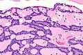
| Image = Canalicular_adenoma_--_high_mag.jpg | |||
| Width = | |||
| Caption = Canalicular adenoma. [[H&E stain]]. (WC) | |||
| Synonyms = | |||
| Micro = cords of tumour ("canals") with beading (characteristic), cystic spaces/tubules, | |||
intraluminal squamous balls (common) | |||
| Subtypes = | |||
| LMDDx = | |||
| Stains = | |||
| IHC = | |||
| EM = | |||
| Molecular = | |||
| IF = | |||
| Gross = | |||
| Grossing = | |||
| Staging = | |||
| Site = [[salivary gland]] - usually upper lip or buccal mucosa | |||
| Assdx = | |||
| Syndromes = | |||
| Clinicalhx = | |||
| Signs = mass lesion | |||
| Symptoms = | |||
| Prevalence = very rare | |||
| Bloodwork = | |||
| Rads = | |||
| Endoscopy = | |||
| Prognosis = | |||
| Other = | |||
| ClinDDx = | |||
| Tx = | |||
}} | |||
'''Canalicular adenoma''' is a rare [[salivary gland]] tumour. | |||
==General== | |||
*Exclusively oral cavity. | |||
Clinical: | |||
*Mass lesion.<ref name=pmid25141970>{{Cite journal | last1 = Thompson | first1 = LD. | last2 = Bauer | first2 = JL. | last3 = Chiosea | first3 = S. | last4 = McHugh | first4 = JB. | last5 = Seethala | first5 = RR. | last6 = Miettinen | first6 = M. | last7 = Müller | first7 = S. | title = Canalicular adenoma: a clinicopathologic and immunohistochemical analysis of 67 cases with a review of the literature. | journal = Head Neck Pathol | volume = 9 | issue = 2 | pages = 181-95 | month = Jun | year = 2015 | doi = 10.1007/s12105-014-0560-6 | PMID = 25141970 }}</ref> | |||
==Gross== | |||
*Classically upper lip - may be buccal mucosa or palate. | |||
Note: | |||
*In one large series of 67 cases:<ref name=pmid25141970/> | |||
**Upper lip 69% (47/67). | |||
**Buccal mucosa 25% (17/67). | |||
**Palate 6% (4/67). | |||
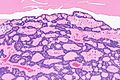
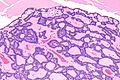
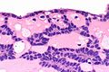
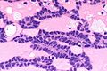
==Microscopic== | |||
Features:<ref name=pmid25141970/> | |||
*Cords of tumour ("canals") with beading - characteristic. | |||
*Cystic spaces/tubules. | |||
*Intraluminal squamous balls - common (~60% of cases). | |||
*Myxoid/bluish stroma. | |||
DDx: | |||
*[[Basal cell adenoma]]. | |||
===Images=== | |||
<gallery> | |||
Image: Canalicular adenoma -- intermed mag.jpg | CA - intermed. mag. (WC) | |||
Image: Canalicular adenoma - alt -- intermed mag.jpg | CA - intermed. mag. (WC) | |||
Image: Canalicular adenoma -- high mag.jpg | CA - high mag. (WC) | |||
Image: Canalicular adenoma -- very high mag.jpg | CA - very high mag. (WC) | |||
Image: Canalicular adenoma - alt -- very high mag.jpg | CA - very high mag. (WC) | |||
</gallery> | |||
====www==== | |||
*[http://www.ncbi.nlm.nih.gov/pmc/articles/PMC4424207/figure/Fig2/ CA (nih.gov)].<ref name=pmid25141970/> | |||
*[http://www.ncbi.nlm.nih.gov/pmc/articles/PMC4424207/figure/Fig3/ CA with myxoid stroma (nih.gov)]. | |||
==IHC== | |||
Features:<ref name=pmid25141970/> | |||
*[[S-100]] +ve - diffuse and strong. | |||
*[[Pankeratin]] +ve - diffuse and strong. | |||
*GFAP +ve - luminal. | |||
*SOX10 +ve - nuclear. | |||
*p16 +ve - luminal squamous balls. | |||
*CK5/6 +ve - luminal squamous balls. | |||
*p63 -ve. | |||
**Nuclei negative, cytoplasm positive. | |||
**Positive in basal cell adenoma. | |||
==See also== | |||
*[[Salivary gland]]. | |||
==References== | |||
{{Reflist|1}} | |||
[[Category:Diagnosis]] | [[Category:Diagnosis]] | ||
[[Category:Head and neck pathology]] | |||
Latest revision as of 21:43, 1 August 2016
| Canalicular adenoma | |
|---|---|
| Diagnosis in short | |
 Canalicular adenoma. H&E stain. (WC) | |
|
| |
| LM |
cords of tumour ("canals") with beading (characteristic), cystic spaces/tubules, intraluminal squamous balls (common) |
| Site | salivary gland - usually upper lip or buccal mucosa |
|
| |
| Signs | mass lesion |
| Prevalence | very rare |
Canalicular adenoma is a rare salivary gland tumour.
General
- Exclusively oral cavity.
Clinical:
- Mass lesion.[1]
Gross
- Classically upper lip - may be buccal mucosa or palate.
Note:
- In one large series of 67 cases:[1]
- Upper lip 69% (47/67).
- Buccal mucosa 25% (17/67).
- Palate 6% (4/67).
Microscopic
Features:[1]
- Cords of tumour ("canals") with beading - characteristic.
- Cystic spaces/tubules.
- Intraluminal squamous balls - common (~60% of cases).
- Myxoid/bluish stroma.
DDx:
Images
CA - intermed. mag. (WC)
CA - intermed. mag. (WC)
CA - high mag. (WC)
CA - very high mag. (WC)
CA - very high mag. (WC)


www
IHC
Features:[1]
- S-100 +ve - diffuse and strong.
- Pankeratin +ve - diffuse and strong.
- GFAP +ve - luminal.
- SOX10 +ve - nuclear.
- p16 +ve - luminal squamous balls.
- CK5/6 +ve - luminal squamous balls.
- p63 -ve.
- Nuclei negative, cytoplasm positive.
- Positive in basal cell adenoma.
See also
References
- ↑ 1.0 1.1 1.2 1.3 1.4 Thompson, LD.; Bauer, JL.; Chiosea, S.; McHugh, JB.; Seethala, RR.; Miettinen, M.; Müller, S. (Jun 2015). "Canalicular adenoma: a clinicopathologic and immunohistochemical analysis of 67 cases with a review of the literature.". Head Neck Pathol 9 (2): 181-95. doi:10.1007/s12105-014-0560-6. PMID 25141970.