Difference between revisions of "Bronchiectasis"
Jump to navigation
Jump to search
(redirect w/ cat.) |
(tweak) |
||
| Line 1: | Line 1: | ||
'''Bronchiectasis''' is a relatively common [[medical lung disease]] with a number of underlying causes. | |||
==General== | |||
*Benign. | |||
*Uncommon. | |||
*Predisposes for infection.<ref name=Ref_PBoD8_693>{{Ref PBoD8|693}}</ref> | |||
**Usually a mixed flora. | |||
**May be predominantly fungal, e.g. ''allergic bronchopulmonary [[aspergillosis]] (ABPA)''. | |||
*Multitude of causes - including: | |||
**[[Cystic fibrosis]] - typically diffusely involvement, unlike other causes.<ref>URL: [http://library.med.utah.edu/WebPath/LUNGHTML/LUNG053.html http://library.med.utah.edu/WebPath/LUNGHTML/LUNG053.html]. Accessed on: 21 February 2012.</ref> | |||
**[[Primary ciliary dyskinesia]]. | |||
==Gross== | |||
*Large airways at the periphery of the lung. | |||
*Central airways larger than the adjacent arteries. | |||
*Typically focal. | |||
Radiologic: | |||
*Central airways larger than the adjacent arteries. | |||
*Airway wall-thickening.<ref>{{Cite journal | last1 = Stockley | first1 = RA. | title = Commentary: bronchiectasis and inflammatory bowel disease. | journal = Thorax | volume = 53 | issue = 6 | pages = 526-7 | month = Jun | year = 1998 | doi = | PMID = 9713456 }}</ref> | |||
*"Tree-in-bud" abnormalities. | |||
===Images=== | |||
<gallery> | |||
Image:Bronchiectasis.jpg | Bronchiectasis. (WC) | |||
</gallery> | |||
www: | |||
*[http://library.med.utah.edu/WebPath/LUNGHTML/LUNG053.html Bronchiectasis (utah.edu)]. | |||
==Microscopic== | |||
Features: | |||
*Dilated airways. | |||
**Airways larger than arteries. | |||
===Image=== | |||
www: | |||
*[http://library.med.utah.edu/WebPath/LUNGHTML/LUNG054.html Bronchiectasis (utah.edu)]. | |||
==See also== | |||
*[[Medical lung diseases]]. | |||
*[[Diffuse panbronchiolitis]]. | |||
==References== | |||
{{Reflist|1}} | |||
[[Category:Diagnosis]] | [[Category:Diagnosis]] | ||
[[Category:Medical lung diseases]] | |||
Latest revision as of 03:25, 18 April 2016
Bronchiectasis is a relatively common medical lung disease with a number of underlying causes.
General
- Benign.
- Uncommon.
- Predisposes for infection.[1]
- Usually a mixed flora.
- May be predominantly fungal, e.g. allergic bronchopulmonary aspergillosis (ABPA).
- Multitude of causes - including:
- Cystic fibrosis - typically diffusely involvement, unlike other causes.[2]
- Primary ciliary dyskinesia.
Gross
- Large airways at the periphery of the lung.
- Central airways larger than the adjacent arteries.
- Typically focal.
Radiologic:
- Central airways larger than the adjacent arteries.
- Airway wall-thickening.[3]
- "Tree-in-bud" abnormalities.
Images
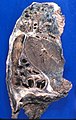
Bronchiectasis. (WC)

www:
Microscopic
Features:
- Dilated airways.
- Airways larger than arteries.
Image
www:
See also
References
- ↑ Kumar, Vinay; Abbas, Abul K.; Fausto, Nelson; Aster, Jon (2009). Robbins and Cotran pathologic basis of disease (8th ed.). Elsevier Saunders. pp. 693. ISBN 978-1416031215.
- ↑ URL: http://library.med.utah.edu/WebPath/LUNGHTML/LUNG053.html. Accessed on: 21 February 2012.
- ↑ Stockley, RA. (Jun 1998). "Commentary: bronchiectasis and inflammatory bowel disease.". Thorax 53 (6): 526-7. PMID 9713456.