Breast calcifications
Jump to navigation
Jump to search
The printable version is no longer supported and may have rendering errors. Please update your browser bookmarks and please use the default browser print function instead.

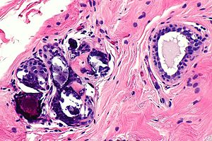
Calcification in benign breast tissue. H&E stain. (WC/Nephron)
Breast calcifications may be found in benign or malignant breast specimens.
General
- Abnormal breast calcifications are considered a marker of malignancy.
- Radiologists can pick-up calcifications that are approximately 100 micrometers; if "calcs" is on the requisition, the pathologist should be finding calcifications this size.[1]
- The large calcifications seen on radiology are approximately 1/5 - 1/6 the size of a HPF, if the field of view (FOV) is ~0.55 mm (as is the case with 22 mm-10x eye pieces and a 40x objective).
Types:
- Calcium phosphate - typically purple.
- Q. How to remember? A. Purple = Phosphate.
- Calcium oxalate - not associated with malignancy.[2]
Microscopic
Features of calcification:
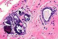
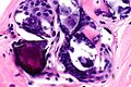
- Purple globs (with concentric rings) on H&E - represent calcium phosphate.
- Often in the lumen of a gland, may be in the stroma.
- Calcific material typically has a well-demarcated border +/- "sharp corners".
Note:
- Calcium oxalate - visible with (light) polarization.
Images

CBBT - intermed. mag.
CBBT - high mag.
CBBT - very high mag.

