Difference between revisions of "Breast calcifications"
Jump to navigation
Jump to search
(more formal) |
|||
| Line 4: | Line 4: | ||
==General== | ==General== | ||
*Abnormal breast calcifications are considered a marker of [[malignancy]]. | *Abnormal breast calcifications are considered a marker of [[malignancy]]. | ||
*Radiologists can pick-up calcifications that are approximately 100 micrometers; if "calcs" is on the requisition | *Radiologists can pick-up calcifications that are approximately 100 micrometers; if "calcs" is on the requisition, the pathologist should be finding calcifications this size.<ref>MUA. 1 October 2010.</ref> | ||
**The large calcifications seen on radiology are approximately 1/5 - 1/6 the size of a HPF, if the field of view (FOV) is ~0.55 mm (as is the case with 22 mm-10x eye pieces and a 40x objective). | **The large calcifications seen on radiology are approximately 1/5 - 1/6 the size of a HPF, if the field of view (FOV) is ~0.55 mm (as is the case with 22 mm-10x eye pieces and a 40x objective). | ||
| Line 10: | Line 10: | ||
*Calcium phosphate - typically purple. | *Calcium phosphate - typically purple. | ||
**Q. How to remember? A. '''P'''urple = '''P'''hosphate. | **Q. How to remember? A. '''P'''urple = '''P'''hosphate. | ||
*Calcium oxalate - not associated with malignancy.<ref name=pmid26769216>{{Cite journal | last1 = Sharma | first1 = T. | last2 = Radosevich | first2 = JA. | last3 = Pachori | first3 = G. | last4 = Mandal | first4 = CC. | title = A Molecular View of Pathological Microcalcification in Breast Cancer. | journal = J Mammary Gland Biol Neoplasia | volume = | issue = | pages = | month = Jan | year = 2016 | doi = 10.1007/s10911-015-9349-9 | PMID = 26769216 }}</ref> | *Calcium oxalate - not associated with malignancy.<ref name=pmid26769216>{{Cite journal | last1 = Sharma | first1 = T. | last2 = Radosevich | first2 = JA. | last3 = Pachori | first3 = G. | last4 = Mandal | first4 = CC. | title = A Molecular View of Pathological Microcalcification in Breast Cancer. | journal = J Mammary Gland Biol Neoplasia | volume = | issue = | pages = | month = Jan | year = 2016 | doi = 10.1007/s10911-015-9349-9 | PMID = 26769216 }}</ref> | ||
==Microscopic== | ==Microscopic== | ||
Latest revision as of 17:10, 5 February 2017

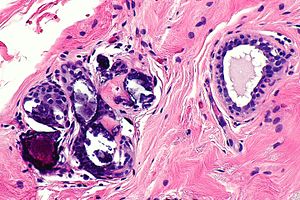
Calcification in benign breast tissue. H&E stain. (WC/Nephron)
Breast calcifications may be found in benign or malignant breast specimens.
General
- Abnormal breast calcifications are considered a marker of malignancy.
- Radiologists can pick-up calcifications that are approximately 100 micrometers; if "calcs" is on the requisition, the pathologist should be finding calcifications this size.[1]
- The large calcifications seen on radiology are approximately 1/5 - 1/6 the size of a HPF, if the field of view (FOV) is ~0.55 mm (as is the case with 22 mm-10x eye pieces and a 40x objective).
Types:
- Calcium phosphate - typically purple.
- Q. How to remember? A. Purple = Phosphate.
- Calcium oxalate - not associated with malignancy.[2]
Microscopic
Features of calcification:
- Purple globs (with concentric rings) on H&E - represent calcium phosphate.
- Often in the lumen of a gland, may be in the stroma.
- Calcific material typically has a well-demarcated border +/- "sharp corners".
Note:
- Calcium oxalate - visible with (light) polarization.
Images

CBBT - intermed. mag.
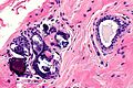
CBBT - high mag.
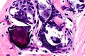
CBBT - very high mag.

