Atrophy of the prostate gland
Jump to navigation
Jump to search
The printable version is no longer supported and may have rendering errors. Please update your browser bookmarks and please use the default browser print function instead.
| Atrophy of the prostate gland | |
|---|---|
| Diagnosis in short | |
 Atrophic prostatic glands. H&E stain. | |
|
| |
| LM | glands typically have a jagged edges/prows (in cancer the glands tend to have round edges), gland density is usually lower than in prostate carcinoma (glands are not back-to-back), nuclei small & hyperchromatic, scant cytoplasm |
| LM DDx | prostate carcinoma - esp. atrophic prostate carcinoma, atypical small acinar proliferation |
| IHC | AMACR -ve, p63 +ve (basal cells), CK34betaE12 +ve (basal cells) |
| Site | prostate gland |
|
| |
| Symptoms | none |
| Prevalence | very common |
| Prognosis | benign |
| Treatment | none |
Atrophy of the prostate gland, also prostatic atrophy, is a common change in the prostate gland.
General
- Considered to be the most common mimicker of prostate carcinoma.[1]
- Small glands - may mimic Gleason pattern 3.
- Inflammatory atrophy seems to be related to HGPIN and prostate cancer;[2] however, the epidemiology is not compelling that this is a significant (clinical) association.[3]
Classification
It can be classified into:[4]
- Focal prostatic atrophy.
- Diffuse prostatic atrophy.
Focal atrophy can be subclassified as:[4]
- Partial.
- Complete.
- Combined.
Microscopic
Features:
- Glands often have a jagged edges/prows (in cancer the glands tend to have round edges) - key feature.
- Prow = forward most part of a ship's bow that cuts through the water.[5]
- You may have come across prow in the context of breast cancer, i.e. tubular carcinoma.
- Prow = forward most part of a ship's bow that cuts through the water.[5]
- Gland density is usually lower than in prostate carcinoma, i.e. glands are not back-to-back - key feature.
- Atrophic glands are often hyperchromatic.[6]
- Scant cytoplasm - usually.
Negatives:
- Nuclei like normal, i.e. nucleoli uncommon.
- Should have two cell layers, i.e. epithelial and myoepithelial (may be difficult to see).
Notes:
- Atrophic glands may be scattered with non-atrophic ones.
- IHC may be misleading - basal cell loss.
DDx:
- Atrophic prostate carcinoma.
- Atypical small acinar proliferation.
- Prostate carcinoma - focal, low grade.
Atrophy versus cancer
| Histologic feature | Atrophy | Cancer |
|---|---|---|
| Glandular architecture/ arrangement |
angulated glands, may look like they originate from one large duct |
round glands, often back-to-back |
| Nuclear hyperchromasia |
marked | moderate |
| Cytoplasm | scant/minimal | moderate, may be amphophilic |
| Basal cells | may be visible | absent |
| Nucleoli | absent | present |
| Secretions in glands |
no | yes - eosinophilic or blue |
Images
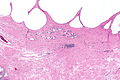
APG - low mag. (WC)
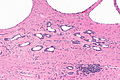
APG - intermed. mag. (WC)
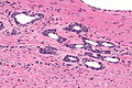
APG - high mag. (WC)
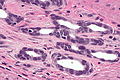
APG - very high mag. (WC)


www:
- Atrophy (webpathology.com).
- Partial atrophy (webpathology.com).
- Sclerotic atrophy (webpathology.com).
IHC
- Classically like normal prostate (AMACR -ve, p63 +ve, CK34betaE12 +ve).
- May be negative for basal cell markers, i.e. p63 and CK34betaE12.[1]
Sign out
Generally, this finding is not reported; it is considered a normal finding.
Left Apex:
- Benign prostate tissue with glandular atrophy.
Micro
The core shows rare, spaced, atrophic appearing glands, mostly with a wavy border and a decreased quantity of cytoplasm. Prominent nucleoli and significant nuclear enlargement are not identified.
There are none of the following: mitoses, adjacent PIN, suspicious luminal material, nuclear hyperchromasia.
See also
References
- ↑ 1.0 1.1 Wang, W.; Sun, X.; Epstein, JI. (Jun 2008). "Partial atrophy on prostate needle biopsy cores: a morphologic and immunohistochemical study.". Am J Surg Pathol 32 (6): 851-7. doi:10.1097/PAS.0b013e31815a0508. PMID 18408595. Cite error: Invalid
<ref>tag; name "pmid18408595" defined multiple times with different content - ↑ De Marzo, AM.; Marchi, VL.; Epstein, JI.; Nelson, WG. (Dec 1999). "Proliferative inflammatory atrophy of the prostate: implications for prostatic carcinogenesis.". Am J Pathol 155 (6): 1985-92. doi:10.1016/S0002-9440(10)65517-4. PMID 10595928.
- ↑ Celma, A.; Servián, P.; Planas, J.; Placer, J.; Quilez, MT.; Arbós, MA.; de Torres, I.; Morote, J. (Mar 2014). "Clinical Significance of Proliferative Inflammatory Atrophy in Prostate Biopsy.". Actas Urol Esp 38 (2): 122-126. doi:10.1016/j.acuro.2013.04.008. PMID 24129226.
- ↑ 4.0 4.1 Billis, A.. "Prostatic atrophy. Clinicopathological significance.". Int Braz J Urol 36 (4): 401-9. PMID 20815946.
- ↑ http://en.wikipedia.org/wiki/Prow
- ↑ SN. June 3, 2009.