Difference between revisions of "Aspergillosis"
Jump to navigation
Jump to search

(redirect) |
(→Stains) |
||
| (3 intermediate revisions by the same user not shown) | |||
| Line 1: | Line 1: | ||
[[Image:Aspergillus - add - very high mag.jpg|thumb|right|Aspergillus with classic fruiting heads. [[H&E stain]].]] | |||
'''Aspergillosis''' is a [[fungi|fungal infection]] due to ''Asperillus''. | |||
==General== | |||
*Due to ''Aspergillus''. | |||
*Fungus. | |||
*Associated with immunosuppression/immunodeficiency. | |||
**Rarely in immune competent individuals.<ref name=pmid7844909>{{Cite journal | last1 = Sugimura | first1 = S. | last2 = Yoshida | first2 = K. | last3 = Oba | first3 = H. | last4 = Hashiguchi | first4 = K. | last5 = Nakajima | first5 = M. | last6 = Moriya | first6 = O. | last7 = Okimoto | first7 = N. | last8 = Niki | first8 = Y. | last9 = Soejima | first9 = R. | title = [Two cases of invasive pulmonary aspergillosis in non-immunocompromised hosts]. | journal = Nihon Kyobu Shikkan Gakkai Zasshi | volume = 32 | issue = 10 | pages = 1032-7 | month = Oct | year = 1994 | doi = | PMID = 7844909 }} | |||
</ref> | |||
==Microscopic== | |||
Features: | |||
*Hyphae that branching with 45 degrees angle - '''key feature'''.<ref name=Ref_APBR682>{{Ref APBR|682}}</ref> | |||
**Uniform width - typically ~3-5 μm. | |||
*Septated - often difficult to see. | |||
*"Fruiting heads" when aerobic - uncommon. | |||
**Spherical structures ~50 micrometres in diameter with radially arranged structures (like spokes of a wheel) +/- an empty centre in the plane of section. | |||
DDx: | |||
*[[Mucormycosis]] - irregular width. | |||
*Scedosporium prolificans - in immunoincompetent individuals.<ref>URL: [http://path.upmc.edu/cases/case290.html http://path.upmc.edu/cases/case290.html]. Accessed on: 14 January 2012.</ref> | |||
*[[Candida]].<ref name=pmid21482725>{{Cite journal | last1 = Guarner | first1 = J. | last2 = Brandt | first2 = ME. | title = Histopathologic diagnosis of fungal infections in the 21st century. | journal = Clin Microbiol Rev | volume = 24 | issue = 2 | pages = 247-80 | month = Apr | year = 2011 | doi = 10.1128/CMR.00053-10 | PMID = 21482725 }}</ref> | |||
====Images==== | |||
<gallery> | |||
Image:Pulmonary_aspergillosis.jpg | Aspergillus (WC) | |||
Image:Pulmonary_aspergillosis_cytology.jpg | Aspergillus - [[cytology]]. (WC) | |||
Image:Aspergillus_-_high_mag.jpg | Aspergillus with fruiting head - high mag. (WC) | |||
Image:Aspergillus_-_add_-_very_high_mag.jpg | Aspergillus with fruiting head - very high mag. (WC) | |||
File:Aspergillosis, angioinvasive, - GMS stain (5390967417).jpg | Angionvasive aspergillosis. (WC/Yale Rosen) | |||
File:Aspergillosis, granulomatous (5390380567).jpg | Granulomatous reaction in aspergillosis - HE.(WC/Yale Rosen) | |||
File:Aspergilloma (5390379559).jpg | Aspergilloma. (WC/Yale Rosen) | |||
</gallery> | |||
www: | |||
*[http://www.ispub.com/journal/the-internet-journal-of-otorhinolaryngology/volume-6-number-1/maxillary-sinus-mycetoma-due-to-aspergillus-niger.article-g03.fs.jpg Aspergillosis - fruiting head (ispub.com)].<ref>URL: [http://www.ispub.com/journal/the-internet-journal-of-otorhinolaryngology/volume-6-number-1/maxillary-sinus-mycetoma-due-to-aspergillus-niger.html http://www.ispub.com/journal/the-internet-journal-of-otorhinolaryngology/volume-6-number-1/maxillary-sinus-mycetoma-due-to-aspergillus-niger.html]. Accessed on: 27 February 2012.</ref> | |||
==Stains== | |||
*[[PAS-D stain]] +ve. | |||
==See also== | |||
*[[Fungi]]. | |||
*[[Microorganisms]]. | |||
==References== | |||
{{Reflist|1}} | |||
[[Category:Diagnosis]] | |||
[[Category:Microorganisms]] | |||
Latest revision as of 20:53, 27 May 2016

Aspergillus with classic fruiting heads. H&E stain.
Aspergillosis is a fungal infection due to Asperillus.
General
- Due to Aspergillus.
- Fungus.
- Associated with immunosuppression/immunodeficiency.
- Rarely in immune competent individuals.[1]
Microscopic
Features:
- Hyphae that branching with 45 degrees angle - key feature.[2]
- Uniform width - typically ~3-5 μm.
- Septated - often difficult to see.
- "Fruiting heads" when aerobic - uncommon.
- Spherical structures ~50 micrometres in diameter with radially arranged structures (like spokes of a wheel) +/- an empty centre in the plane of section.
DDx:
- Mucormycosis - irregular width.
- Scedosporium prolificans - in immunoincompetent individuals.[3]
- Candida.[4]
Images
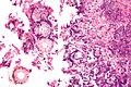
Aspergillus (WC)

Aspergillus - cytology. (WC)
Aspergillus with fruiting head - high mag. (WC)
Aspergillus with fruiting head - very high mag. (WC)
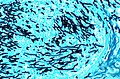
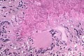
Angionvasive aspergillosis. (WC/Yale Rosen)
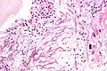
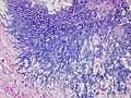
Granulomatous reaction in aspergillosis - HE.(WC/Yale Rosen)
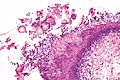
Aspergilloma. (WC/Yale Rosen)


.jpg)
.jpg)
.jpg)
www:
Stains
- PAS-D stain +ve.
See also
References
- ↑ Sugimura, S.; Yoshida, K.; Oba, H.; Hashiguchi, K.; Nakajima, M.; Moriya, O.; Okimoto, N.; Niki, Y. et al. (Oct 1994). "[Two cases of invasive pulmonary aspergillosis in non-immunocompromised hosts].". Nihon Kyobu Shikkan Gakkai Zasshi 32 (10): 1032-7. PMID 7844909.
- ↑ Lefkowitch, Jay H. (2006). Anatomic Pathology Board Review (1st ed.). Saunders. pp. 682. ISBN 978-1416025887.
- ↑ URL: http://path.upmc.edu/cases/case290.html. Accessed on: 14 January 2012.
- ↑ Guarner, J.; Brandt, ME. (Apr 2011). "Histopathologic diagnosis of fungal infections in the 21st century.". Clin Microbiol Rev 24 (2): 247-80. doi:10.1128/CMR.00053-10. PMID 21482725.
- ↑ URL: http://www.ispub.com/journal/the-internet-journal-of-otorhinolaryngology/volume-6-number-1/maxillary-sinus-mycetoma-due-to-aspergillus-niger.html. Accessed on: 27 February 2012.