Difference between revisions of "Angiomyolipoma"
(→Images) |
(+may involve LNs) |
||
| (21 intermediate revisions by 2 users not shown) | |||
| Line 1: | Line 1: | ||
{{ Infobox diagnosis | {{ Infobox diagnosis | ||
| Name = {{PAGENAME}} | | Name = {{PAGENAME}} | ||
| Image = | | Image = Angiomyolipoma -- low mag.jpg | ||
| Width = | | Width = | ||
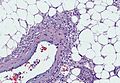
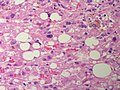
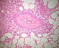
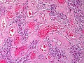
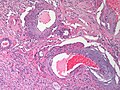
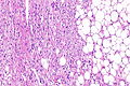
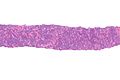
| Caption = | | Caption = [[Micrograph]] of an angiomyolipoma - showing the three components (blood vessels (''angio-'') - left top, muscle (''myo-'') - elsewhere, fat (''lipoma'') - right bottom). [[H&E stain]]. | ||
| Synonyms = | | Synonyms = | ||
| Micro = smooth muscle, adipose tissue (not always present), abundant blood vessels | | Micro = smooth muscle, adipose tissue (not always present), abundant blood vessels | ||
| Line 38: | Line 38: | ||
*Presentations: flank pain, hematuria, incidentaloma.<ref name=pmid18805573>{{Cite journal | last1 = Seyam | first1 = RM. | last2 = Bissada | first2 = NK. | last3 = Kattan | first3 = SA. | last4 = Mokhtar | first4 = AA. | last5 = Aslam | first5 = M. | last6 = Fahmy | first6 = WE. | last7 = Mourad | first7 = WA. | last8 = Binmahfouz | first8 = AA. | last9 = Alzahrani | first9 = HM. | title = Changing trends in presentation, diagnosis and management of renal angiomyolipoma: comparison of sporadic and tuberous sclerosis complex-associated forms. | journal = Urology | volume = 72 | issue = 5 | pages = 1077-82 | month = Nov | year = 2008 | doi = 10.1016/j.urology.2008.07.049 | PMID = 18805573 }}</ref> | *Presentations: flank pain, hematuria, incidentaloma.<ref name=pmid18805573>{{Cite journal | last1 = Seyam | first1 = RM. | last2 = Bissada | first2 = NK. | last3 = Kattan | first3 = SA. | last4 = Mokhtar | first4 = AA. | last5 = Aslam | first5 = M. | last6 = Fahmy | first6 = WE. | last7 = Mourad | first7 = WA. | last8 = Binmahfouz | first8 = AA. | last9 = Alzahrani | first9 = HM. | title = Changing trends in presentation, diagnosis and management of renal angiomyolipoma: comparison of sporadic and tuberous sclerosis complex-associated forms. | journal = Urology | volume = 72 | issue = 5 | pages = 1077-82 | month = Nov | year = 2008 | doi = 10.1016/j.urology.2008.07.049 | PMID = 18805573 }}</ref> | ||
**Tumours >4 cm considered a risk for bleeding.<ref name=pmid21571778>{{Cite journal | last1 = Abrams | first1 = J. | last2 = Yee | first2 = DC. | last3 = Clark | first3 = TW. | title = Transradial embolization of a bleeding renal angiomyolipoma. | journal = Vasc Endovascular Surg | volume = 45 | issue = 5 | pages = 470-3 | month = Jul | year = 2011 | doi = 10.1177/1538574411408352 | PMID = 21571778 }}</ref> | **Tumours >4 cm considered a risk for bleeding.<ref name=pmid21571778>{{Cite journal | last1 = Abrams | first1 = J. | last2 = Yee | first2 = DC. | last3 = Clark | first3 = TW. | title = Transradial embolization of a bleeding renal angiomyolipoma. | journal = Vasc Endovascular Surg | volume = 45 | issue = 5 | pages = 470-3 | month = Jul | year = 2011 | doi = 10.1177/1538574411408352 | PMID = 21571778 }}</ref> | ||
**May bleed spontaneously ([[Wunderlich's syndrome]]).<ref name=pmid19104113>{{Cite journal | last1 = Moratalla | first1 = MB. | title = Wunderlich's syndrome due to spontaneous rupture of large bilateral angiomyolipomas. | journal = Emerg Med J | volume = 26 | issue = 1 | pages = 72 | month = Jan | year = 2009 | doi = 10.1136/emj.2008.062091 | PMID = 19104113 }}</ref> | |||
*AMLs occur may be elsewhere in the body, e.g. liver,<ref name=pmid15498214>{{Cite journal | last1 = Zhang | first1 = SH. | last2 = Cong | first2 = WM. | last3 = Xian | first3 = ZH. | last4 = Wu | first4 = WQ. | last5 = Dong | first5 = H. | last6 = Wu | first6 = MC. | title = [Morphologic variants and immunohistochemical features of hepatic angiomyolipoma.] | journal = Zhonghua Bing Li Xue Za Zhi | volume = 33 | issue = 5 | pages = 437-40 | month = Oct | year = 2004 | doi = | PMID = 15498214 }} | *AMLs occur may be elsewhere in the body, e.g. liver,<ref name=pmid15498214>{{Cite journal | last1 = Zhang | first1 = SH. | last2 = Cong | first2 = WM. | last3 = Xian | first3 = ZH. | last4 = Wu | first4 = WQ. | last5 = Dong | first5 = H. | last6 = Wu | first6 = MC. | title = [Morphologic variants and immunohistochemical features of hepatic angiomyolipoma.] | journal = Zhonghua Bing Li Xue Za Zhi | volume = 33 | issue = 5 | pages = 437-40 | month = Oct | year = 2004 | doi = | PMID = 15498214 }} | ||
</ref> but are most common in the periadrenal and perirenal fat. | </ref> but are most common in the periadrenal and perirenal fat. | ||
| Line 51: | Line 52: | ||
**This is not confirmed by all studies.<ref name=pmid18852677>{{Cite journal | last1 = Aydin | first1 = H. | last2 = Magi-Galluzzi | first2 = C. | last3 = Lane | first3 = BR. | last4 = Sercia | first4 = L. | last5 = Lopez | first5 = JI. | last6 = Rini | first6 = BI. | last7 = Zhou | first7 = M. | title = Renal angiomyolipoma: clinicopathologic study of 194 cases with emphasis on the epithelioid histology and tuberous sclerosis association. | journal = Am J Surg Pathol | volume = 33 | issue = 2 | pages = 289-97 | month = Feb | year = 2009 | doi = 10.1097/PAS.0b013e31817ed7a6 | PMID = 18852677 }}</ref> | **This is not confirmed by all studies.<ref name=pmid18852677>{{Cite journal | last1 = Aydin | first1 = H. | last2 = Magi-Galluzzi | first2 = C. | last3 = Lane | first3 = BR. | last4 = Sercia | first4 = L. | last5 = Lopez | first5 = JI. | last6 = Rini | first6 = BI. | last7 = Zhou | first7 = M. | title = Renal angiomyolipoma: clinicopathologic study of 194 cases with emphasis on the epithelioid histology and tuberous sclerosis association. | journal = Am J Surg Pathol | volume = 33 | issue = 2 | pages = 289-97 | month = Feb | year = 2009 | doi = 10.1097/PAS.0b013e31817ed7a6 | PMID = 18852677 }}</ref> | ||
*More common in women than men - both in sporadic AMLs and those associated with tuberous sclerosis.<ref name=pmid17003820>{{cite journal |author=Rakowski SK, Winterkorn EB, Paul E, Steele DJ, Halpern EF, Thiele EA |title=Renal manifestations of tuberous sclerosis complex: Incidence, prognosis, and predictive factors |journal=Kidney Int. |volume=70 |issue=10 |pages=1777–82 |year=2006 |month=November |pmid=17003820 |doi=10.1038/sj.ki.5001853 |url=}}</ref> | *More common in women than men - both in sporadic AMLs and those associated with tuberous sclerosis.<ref name=pmid17003820>{{cite journal |author=Rakowski SK, Winterkorn EB, Paul E, Steele DJ, Halpern EF, Thiele EA |title=Renal manifestations of tuberous sclerosis complex: Incidence, prognosis, and predictive factors |journal=Kidney Int. |volume=70 |issue=10 |pages=1777–82 |year=2006 |month=November |pmid=17003820 |doi=10.1038/sj.ki.5001853 |url=}}</ref> | ||
==Gross== | |||
*Well circumscribed - uniform yellow. | |||
Note: | |||
*Small resected AML are often fat poor. | |||
*May resemble a renal cell carcinoma on gross. | |||
Radiology: | |||
*<=10 HU in an intermediate size tumour.<ref name=pmid21555349>{{Cite journal | last1 = Davenport | first1 = MS. | last2 = Neville | first2 = AM. | last3 = Ellis | first3 = JH. | last4 = Cohan | first4 = RH. | last5 = Chaudhry | first5 = HS. | last6 = Leder | first6 = RA. | title = Diagnosis of renal angiomyolipoma with hounsfield unit thresholds: effect of size of region of interest and nephrographic phase imaging. | journal = Radiology | volume = 260 | issue = 1 | pages = 158-65 | month = Jul | year = 2011 | doi = 10.1148/radiol.11102476 | PMID = 21555349 }}</ref> | |||
Image: | |||
*[http://webpathology.com/image.asp?n=6&Case=72 AML (webpathology.com)]. | |||
==Microscopic== | ==Microscopic== | ||
| Line 57: | Line 71: | ||
*Adipose tissue - not always present<ref name=pmid15584043>{{Cite journal | last1 = Crapanzano | first1 = JP. | title = Fine-needle aspiration of renal angiomyolipoma: cytological findings and diagnostic pitfalls in a series of five cases. | journal = Diagn Cytopathol | volume = 32 | issue = 1 | pages = 53-7 | month = Jan | year = 2005 | doi = 10.1002/dc.20179 | PMID = 15584043 }}</ref> - '''key feature'''. | *Adipose tissue - not always present<ref name=pmid15584043>{{Cite journal | last1 = Crapanzano | first1 = JP. | title = Fine-needle aspiration of renal angiomyolipoma: cytological findings and diagnostic pitfalls in a series of five cases. | journal = Diagn Cytopathol | volume = 32 | issue = 1 | pages = 53-7 | month = Jan | year = 2005 | doi = 10.1002/dc.20179 | PMID = 15584043 }}</ref> - '''key feature'''. | ||
*Abundant [[blood vessel]]s. | *Abundant [[blood vessel]]s. | ||
Notes: | |||
*May have melanin pigment - occasionally extensive.<ref name=pmid26118214>{{Cite journal | last1 = Reddy | first1 = R. | last2 = Lewin | first2 = JR. | last3 = Shenoy | first3 = V. | title = Pigmented Epithelioid Angiomyolipoma of the Kidney. | journal = J Miss State Med Assoc | volume = 56 | issue = 4 | pages = 92-4 | month = Apr | year = 2015 | doi = | PMID = 26118214 }}</ref><ref name=pmid23136579>{{Cite journal | last1 = Chang | first1 = H. | last2 = Jung | first2 = W. | last3 = Kang | first3 = Y. | last4 = Jung | first4 = WY. | title = Pigmented perivascular epithelioid cell tumor (PEComa) of the kidney: a case report and review of the literature. | journal = Korean J Pathol | volume = 46 | issue = 5 | pages = 499-502 | month = Oct | year = 2012 | doi = 10.4132/KoreanJPathol.2012.46.5.499 | PMID = 23136579 }}</ref> | |||
*May involve lymph nodes - case reports.<ref name=pmid2294869>{{cite journal |authors=Ro JY, Ayala AG, el-Naggar A, Grignon DJ, Hogan SF, Howard DR |title=Angiomyolipoma of kidney with lymph node involvement. DNA flow cytometric analysis |journal=Arch Pathol Lab Med |volume=114 |issue=1 |pages=65–7 |date=January 1990 |pmid=2294869 |doi= |url=}}</ref><ref name=pmid12696187>{{cite journal |vauthor=Shiga Y, Tsutsumi M, Suzuki K, Ishikawa S, Shimogama T |title=Angiomyolipoma with regional lymph node involvement: a case report and literature review |journal=Hinyokika Kiyo |volume=49 |issue=2 |pages=81–6 |date=February 2003 |pmid=12696187 |doi= |url=}}</ref> | |||
DDx: | DDx: | ||
| Line 72: | Line 90: | ||
Image: Adrenal Angiomyolipoma MP CTR.jpg|Angiomyolipoma (SKB) | Image: Adrenal Angiomyolipoma MP CTR.jpg|Angiomyolipoma (SKB) | ||
Image: Kidney Angiomyolipoma Hemorrhage HP CTR.jpg|Renal angiomyolipoma with hemorrhage (SKB) | Image: Kidney Angiomyolipoma Hemorrhage HP CTR.jpg|Renal angiomyolipoma with hemorrhage (SKB) | ||
Image: Kidney Angiomyolipoma Hemorrhage HP2 CTR.jpg|Renal angiomyolipoma with hemorrhage (SKB) | |||
Image: Kidney Angiomyolipoma HP CTR (2).jpg|Renal angiomyolipoma (SKB) | Image: Kidney Angiomyolipoma HP CTR (2).jpg|Renal angiomyolipoma (SKB) | ||
Image: Kidney Angiomyolipoma 4 LP CTR.jpg|Renal angiomyolipoma - low power (SKB) | Image: Kidney Angiomyolipoma 4 LP CTR.jpg|Renal angiomyolipoma - low power (SKB) | ||
| Line 78: | Line 97: | ||
Image: Kidney Angiomyolipoma 4 MP CTR.jpg|Renal angiomyolipoma - medium power (SKB) | Image: Kidney Angiomyolipoma 4 MP CTR.jpg|Renal angiomyolipoma - medium power (SKB) | ||
Image: Kidney Angiomyolipoma Hemorrhage MP CTR.jpg|Renal angiomyolipoma - medium power (SKB) | Image: Kidney Angiomyolipoma Hemorrhage MP CTR.jpg|Renal angiomyolipoma - medium power (SKB) | ||
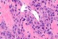
Image: Kidney Angiomyolipoma 1 PA.jpg|Renal angiomyolipoma (SKB) | Image: Kidney Angiomyolipoma 1 PA.jpg|Renal angiomyolipoma - Tumor cells stream off a blood vessel (SKB) | ||
Image: Kidney Angiomyolipoma MP CTR.jpg|Renal Angiomyolipoma - medium power (SKB) | |||
Image: Kidney Angiomyolipoma HP CTR.jpg|Renal angiomyolipoma - high power (SKB) | Image: Kidney Angiomyolipoma HP CTR.jpg|Renal angiomyolipoma - high power (SKB) | ||
Image: Kidney Angiomyolipoma MP2 CTR.jpg|Renal Angiomyolipoma - medium power (SKB) | |||
Image: Kidney Angiomyolipoma MP CTR (2).jpg|Renal Angiomyolipoma - medium power (SKB) | |||
Image: Kidney Angiomyolipoma MP CTR (3).jpg|Renal Angiomyolipoma - medium power (SKB) | |||
Image: Kidney Angiomyolipoma SolidType HP PA.jpg|Renal Angiomyolipoma - solid type - high power (SKB) | |||
Image: Kidney Angiomyolipoma SolidType HP2 PA.jpg|Renal angiomyolipoma - solid type (SKB) | |||
Image: Kidney Angiomyolipoma SolidVariant MP3 CTR.jpg|Renal angiomyolipoma - solid type (SKB) | |||
Image: Kidney Angiomyolipoma SolidVariant MP CTR.jpg|Renal angiomyolipoma - solid type (SKB) | |||
Image: Kidney Angiomyolipoma SolidType HP3 PA.jpg|Renal angiomyolipoma - solid type (SKB) | |||
Image: Kidney Angiomyolipoma SolidVariant MP2 CTR.jpg|Renal angiomyolipoma - solid type (SKB) | |||
</gallery> | |||
====Case==== | |||
<gallery> | |||
Image: Angiomyolipoma -- low mag.jpg | AML - low mag. | |||
Image: Angiomyolipoma -- intermed mag.jpg | AML - intermed. mag. | |||
Image: Angiomyolipoma - alt -- intermed mag.jpg | AML - intermed. mag. | |||
Image: Angiomyolipoma -- high mag.jpg | AML - intermed. mag. | |||
Image: Angiomyolipoma - alt -- high mag.jpg | AML - intermed. mag. | |||
</gallery> | </gallery> | ||
| Line 90: | Line 127: | ||
DDx: | DDx: | ||
*[[Clear cell renal cell carcinoma]] | *[[Clear cell renal cell carcinoma]]. | ||
*[[Xp11.2 translocation carcinoma]]. | *[[Xp11.2 translocation carcinoma]]. | ||
*Renal cell carcinoma with angioleiomyoma-like stroma.<ref name=pmid25189644>{{Cite journal | last1 = Williamson | first1 = SR. | last2 = Cheng | first2 = L. | last3 = Eble | first3 = JN. | last4 = True | first4 = LD. | last5 = Gupta | first5 = NS. | last6 = Wang | first6 = M. | last7 = Zhang | first7 = S. | last8 = Grignon | first8 = DJ. | title = Renal cell carcinoma with angioleiomyoma-like stroma: clinicopathological, immunohistochemical, and molecular features supporting classification as a distinct entity. | journal = Mod Pathol | volume = | issue = | pages = | month = Sep | year = 2014 | doi = 10.1038/modpathol.2014.105 | PMID = 25189644 }}</ref> | *Renal cell carcinoma with angioleiomyoma-like stroma.<ref name=pmid25189644>{{Cite journal | last1 = Williamson | first1 = SR. | last2 = Cheng | first2 = L. | last3 = Eble | first3 = JN. | last4 = True | first4 = LD. | last5 = Gupta | first5 = NS. | last6 = Wang | first6 = M. | last7 = Zhang | first7 = S. | last8 = Grignon | first8 = DJ. | title = Renal cell carcinoma with angioleiomyoma-like stroma: clinicopathological, immunohistochemical, and molecular features supporting classification as a distinct entity. | journal = Mod Pathol | volume = | issue = | pages = | month = Sep | year = 2014 | doi = 10.1038/modpathol.2014.105 | PMID = 25189644 }}</ref> | ||
====Images==== | ====Images==== | ||
www | <gallery> | ||
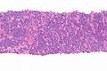
Image: Epithelioid angiomyolipoma -- very low mag.jpg | EMAL - very low mag. | |||
Image: Epithelioid angiomyolipoma -- low mag.jpg | EMAL - low mag. | |||
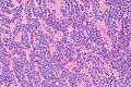
Image: Epithelioid angiomyolipoma -- intermed mag.jpg | EMAL - intermed. mag. | |||
Image: Epithelioid angiomyolipoma -- high mag.jpg | EMAL - high mag. | |||
Image: Epithelioid angiomyolipoma -- very high mag.jpg | EMAL - very high mag. | |||
Image: Epithelioid angiomyolipoma - alt -- low mag.jpg | EMAL - low mag. | |||
Image: Epithelioid angiomyolipoma - alt -- intermed mag.jpg | EMAL - intermed. mag. | |||
</gallery> | |||
=====www===== | |||
*[http://www.nature.com/modpathol/journal/v26/n10/fig_tab/modpathol201372f4.html#figure-title Epithelioid angiomyolipoma (nature.com)].<ref name=pmid23599151>{{Cite journal | last1 = He | first1 = W. | last2 = Cheville | first2 = JC. | last3 = Sadow | first3 = PM. | last4 = Gopalan | first4 = A. | last5 = Fine | first5 = SW. | last6 = Al-Ahmadie | first6 = HA. | last7 = Chen | first7 = YB. | last8 = Oliva | first8 = E. | last9 = Russo | first9 = P. | title = Epithelioid angiomyolipoma of the kidney: pathological features and clinical outcome in a series of consecutively resected tumors. | journal = Mod Pathol | volume = 26 | issue = 10 | pages = 1355-64 | month = Oct | year = 2013 | doi = 10.1038/modpathol.2013.72 | PMID = 23599151 }}</ref> | *[http://www.nature.com/modpathol/journal/v26/n10/fig_tab/modpathol201372f4.html#figure-title Epithelioid angiomyolipoma (nature.com)].<ref name=pmid23599151>{{Cite journal | last1 = He | first1 = W. | last2 = Cheville | first2 = JC. | last3 = Sadow | first3 = PM. | last4 = Gopalan | first4 = A. | last5 = Fine | first5 = SW. | last6 = Al-Ahmadie | first6 = HA. | last7 = Chen | first7 = YB. | last8 = Oliva | first8 = E. | last9 = Russo | first9 = P. | title = Epithelioid angiomyolipoma of the kidney: pathological features and clinical outcome in a series of consecutively resected tumors. | journal = Mod Pathol | volume = 26 | issue = 10 | pages = 1355-64 | month = Oct | year = 2013 | doi = 10.1038/modpathol.2013.72 | PMID = 23599151 }}</ref> | ||
*[http://www.nature.com/modpathol/journal/v26/n10/fig_tab/modpathol201372f2.html#figure-title Epithelioid AML - spectrum of patterns (nature.com)].<ref name=pmid23599151/> | *[http://www.nature.com/modpathol/journal/v26/n10/fig_tab/modpathol201372f2.html#figure-title Epithelioid AML - spectrum of patterns (nature.com)].<ref name=pmid23599151/> | ||
| Line 141: | Line 187: | ||
===Biopsy=== | ===Biopsy=== | ||
<pre> | |||
Left Kidney, Mass Lesion, Core Biopsy: | |||
- Angiomyolipoma, see comment. | |||
Comment: | |||
The tumour stains as follow: | |||
POSITIVE: melan A (focal, rare). | |||
NEGATIVE: PAX8, AE1/AE3, HMB-45, desmin, PR, ER. | |||
</pre> | |||
====Alternate==== | |||
<pre> | <pre> | ||
MASS OF LEFT KIDNEY, CORE BIOPSY: | MASS OF LEFT KIDNEY, CORE BIOPSY: | ||
| Line 152: | Line 209: | ||
====Micro==== | ====Micro==== | ||
The sections show a tumour composed of spindle cells in a | The sections show a tumour composed of spindle cells in a fascicular arrangement with scant adipose tissue and prominent muscular blood vessels. Rare nuclear enlargement is seen. No significant nuclear atypia is appreciated. Mitotic activity is not apparent. No renal parenchyma is identified. | ||
==See also== | ==See also== | ||
Latest revision as of 15:08, 31 March 2023
| Angiomyolipoma | |
|---|---|
| Diagnosis in short | |
 Micrograph of an angiomyolipoma - showing the three components (blood vessels (angio-) - left top, muscle (myo-) - elsewhere, fat (lipoma) - right bottom). H&E stain. | |
|
| |
| LM | smooth muscle, adipose tissue (not always present), abundant blood vessels |
| Subtypes | conventional, epithelioid angiomyolipoma |
| LM DDx | clear cell renal cell carcinoma (esp. for epithelioid variant), Xp11.2 translocation carcinoma (esp. for epithelioid variant), well-differentiated retroperitoneal sarcoma (esp. leiomyosarcoma & liposarcoma), renal cell carcinoma with sarcomatoid differentiation, renal leiomyoma |
| IHC | HMB-45 +ve, Melan A +ve, SMA +ve, PAX8 -ve, CK (pooled) -ve |
| Site | kidney (see kidney tumours), other sites |
|
| |
| Syndromes | tuberous sclerosis |
|
| |
| Prevalence | uncommon |
| Radiology | classically has regions consistent with fat |
| Prognosis | benign, epithelioid variant may be aggressive |
| Clin. DDx | other kidney tumours |
| Treatment | surgery - esp. if large or imaging characteristics ambiguous |
Angiomyolipoma, abbreviated AML, is a benign mesenchymal tumour that is associated with tuberous sclerosis and belongs to the PEComa group of tumours.
It is typically found in the kidney; however, occasionally it is seen extrarenal.[1]
General
- Benign mesenchymal tumour.
- Presentations: flank pain, hematuria, incidentaloma.[2]
- Tumours >4 cm considered a risk for bleeding.[3]
- May bleed spontaneously (Wunderlich's syndrome).[4]
- AMLs occur may be elsewhere in the body, e.g. liver,[5] but are most common in the periadrenal and perirenal fat.
- In the PEComa group of tumours.
Epidemiology
- May be associated with tuberous sclerosis -- 70% have an AML.
- When compared to sporadic cases:
- More often bilateral.
- Usually bigger.
- Reported to often have estrogen and progesterone receptors - in the context of lymphangioleiomyomatosis.[6]
- When compared to sporadic cases:
- There is a suggestion that an epithelioid variant is more worrisome.[7]
- This is not confirmed by all studies.[8]
- More common in women than men - both in sporadic AMLs and those associated with tuberous sclerosis.[9]
Gross
- Well circumscribed - uniform yellow.
Note:
- Small resected AML are often fat poor.
- May resemble a renal cell carcinoma on gross.
Radiology:
- <=10 HU in an intermediate size tumour.[10]
Image:
Microscopic
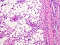
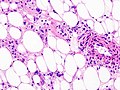
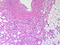
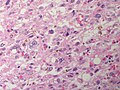
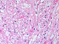
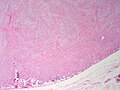
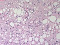
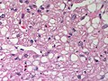
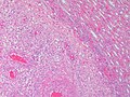
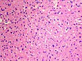
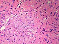
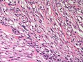
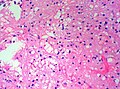
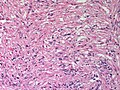
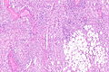
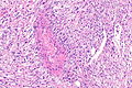
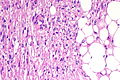
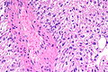
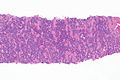
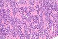
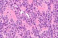
Features:
- Smooth muscle.
- Adipose tissue - not always present[11] - key feature.
- Abundant blood vessels.
Notes:
- May have melanin pigment - occasionally extensive.[12][13]
- May involve lymph nodes - case reports.[14][15]
DDx:
- Retroperitoneal sarcoma.
- Renal cell carcinoma with sarcomatoid differentiation.
- Renal leiomyoma - very rare - desmin +ve, HMB-45 -ve.[16]
Images
AML (WC).
AML (WC/KGH).
AML (WC/KGH).
Angiomyolipoma (SKB)
Renal angiomyolipoma with hemorrhage (SKB)
Renal angiomyolipoma with hemorrhage (SKB)
Renal angiomyolipoma (SKB)
Renal angiomyolipoma - low power (SKB)
Renal angiomyolipoma- medium power (SKB)
Renal angiomyolipoma - low power (SKB)
Renal angiomyolipoma - medium power (SKB)
Renal angiomyolipoma - medium power (SKB)
Renal angiomyolipoma - Tumor cells stream off a blood vessel (SKB)
Renal Angiomyolipoma - medium power (SKB)
Renal angiomyolipoma - high power (SKB)
Renal Angiomyolipoma - medium power (SKB)

Renal Angiomyolipoma - medium power (SKB)
Renal Angiomyolipoma - medium power (SKB)
Renal Angiomyolipoma - solid type - high power (SKB)
Renal angiomyolipoma - solid type (SKB)
Renal angiomyolipoma - solid type (SKB)
Renal angiomyolipoma - solid type (SKB)
Renal angiomyolipoma - solid type (SKB)
Renal angiomyolipoma - solid type (SKB)

.jpg)
.jpg)


.jpg)


.jpg)
.jpg)


Case
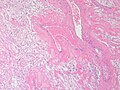
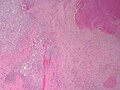
AML - low mag.
AML - intermed. mag.
AML - intermed. mag.
AML - intermed. mag.
AML - intermed. mag.


Epithelioid angiomyolipoma
Features:
- Carcinoma-like morphology.
- +/-Spindle cells.
- "High grade" nuclei.
- Pleomorphic nuclei.
DDx:
- Clear cell renal cell carcinoma.
- Xp11.2 translocation carcinoma.
- Renal cell carcinoma with angioleiomyoma-like stroma.[17]
Images
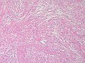
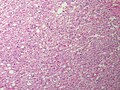
EMAL - very low mag.
EMAL - low mag.
EMAL - intermed. mag.
EMAL - high mag.
EMAL - very high mag.
EMAL - low mag.
EMAL - intermed. mag.


www
- Epithelioid angiomyolipoma (nature.com).[18]
- Epithelioid AML - spectrum of patterns (nature.com).[18]
- Epithelioid AML - variants (nature.com).[18]
- Epithelioid AML (rsna.org).[19]
- Atypical epithelioid AML (archivesofpathology.org).[20]
Cytologic
Features[11]
- Nuclei - round/ovoid.
- Chromatin - bland.
IHC
- Melanocytic markers +ve.[21]
- HMB-45 +ve in all cases (15/15).[22]
- Melan A +ve in ~87% of cases (13/15).
- Epithelial markers -ve[21], e.g. EMA and AE1/AE3.
- SMA +ve.
- CD117 +ve/-ve.
- Ki-67:[23]
- Epithelioid variant of AML +ve.
- Conventional AML -ve.
A panel:
- CK (pooled), Desmin, Melan A, HMB-45.
Epithelioid AML versus Xp11.2 translocation carcinoma
- PAX8 -ve.
- Positive in Xp11.2 translocation carcinoma.
- CAIX -ve.
- AE1/AE3 -ve.
- EMA -ve.
Sign out
RIGHT KIDNEY, PARTIAL NEPHRECTOMY:
- ANGIOMYOLIPOMA.
Micro
The section show a circumscribed tumour composed of muscle-like tissue and adipose tissue with prominent muscular blood vessels. Rare nuclear enlargement is seen. No signficant nuclear atypia is appreciated. Mitotic activity is not apparent.
Biopsy
Left Kidney, Mass Lesion, Core Biopsy:
     - Angiomyolipoma, see comment.

Comment:
The tumour stains as follow:
POSITIVE: melan A (focal, rare).
NEGATIVE: PAX8, AE1/AE3, HMB-45, desmin, PR, ER.
Alternate
MASS OF LEFT KIDNEY, CORE BIOPSY:
- COMPATIBLE WITH ANGIOMYOLIPOMA.

COMMENT:
The tumour stains as follows:
POSITIVE: Melan A, HMB-45, desmin.
NEGATIVE: CK (pooled).
Micro
The sections show a tumour composed of spindle cells in a fascicular arrangement with scant adipose tissue and prominent muscular blood vessels. Rare nuclear enlargement is seen. No significant nuclear atypia is appreciated. Mitotic activity is not apparent. No renal parenchyma is identified.
See also
References
- ↑ Minja, EJ.; Pellerin, M.; Saviano, N.; Chamberlain, RS. (2012). "Retroperitoneal extrarenal angiomyolipomas: an evidence-based approach to a rare clinical entity.". Case Rep Nephrol 2012: 374107. doi:10.1155/2012/374107. PMID 24555133.
- ↑ Seyam, RM.; Bissada, NK.; Kattan, SA.; Mokhtar, AA.; Aslam, M.; Fahmy, WE.; Mourad, WA.; Binmahfouz, AA. et al. (Nov 2008). "Changing trends in presentation, diagnosis and management of renal angiomyolipoma: comparison of sporadic and tuberous sclerosis complex-associated forms.". Urology 72 (5): 1077-82. doi:10.1016/j.urology.2008.07.049. PMID 18805573.
- ↑ Abrams, J.; Yee, DC.; Clark, TW. (Jul 2011). "Transradial embolization of a bleeding renal angiomyolipoma.". Vasc Endovascular Surg 45 (5): 470-3. doi:10.1177/1538574411408352. PMID 21571778.
- ↑ Moratalla, MB. (Jan 2009). "Wunderlich's syndrome due to spontaneous rupture of large bilateral angiomyolipomas.". Emerg Med J 26 (1): 72. doi:10.1136/emj.2008.062091. PMID 19104113.
- ↑ Zhang, SH.; Cong, WM.; Xian, ZH.; Wu, WQ.; Dong, H.; Wu, MC. (Oct 2004). "[Morphologic variants and immunohistochemical features of hepatic angiomyolipoma.]". Zhonghua Bing Li Xue Za Zhi 33 (5): 437-40. PMID 15498214.
- ↑ Logginidou, H.; Ao, X.; Russo, I.; Henske, EP. (Jan 2000). "Frequent estrogen and progesterone receptor immunoreactivity in renal angiomyolipomas from women with pulmonary lymphangioleiomyomatosis.". Chest 117 (1): 25-30. PMID 10631194.
- ↑ Nelson, CP.; Sanda, MG. (Oct 2002). "Contemporary diagnosis and management of renal angiomyolipoma.". J Urol 168 (4 Pt 1): 1315-25. doi:10.1097/01.ju.0000028200.86216.b2. PMID 12352384.
- ↑ Aydin, H.; Magi-Galluzzi, C.; Lane, BR.; Sercia, L.; Lopez, JI.; Rini, BI.; Zhou, M. (Feb 2009). "Renal angiomyolipoma: clinicopathologic study of 194 cases with emphasis on the epithelioid histology and tuberous sclerosis association.". Am J Surg Pathol 33 (2): 289-97. doi:10.1097/PAS.0b013e31817ed7a6. PMID 18852677.
- ↑ Rakowski SK, Winterkorn EB, Paul E, Steele DJ, Halpern EF, Thiele EA (November 2006). "Renal manifestations of tuberous sclerosis complex: Incidence, prognosis, and predictive factors". Kidney Int. 70 (10): 1777–82. doi:10.1038/sj.ki.5001853. PMID 17003820.
- ↑ Davenport, MS.; Neville, AM.; Ellis, JH.; Cohan, RH.; Chaudhry, HS.; Leder, RA. (Jul 2011). "Diagnosis of renal angiomyolipoma with hounsfield unit thresholds: effect of size of region of interest and nephrographic phase imaging.". Radiology 260 (1): 158-65. doi:10.1148/radiol.11102476. PMID 21555349.
- ↑ 11.0 11.1 Crapanzano, JP. (Jan 2005). "Fine-needle aspiration of renal angiomyolipoma: cytological findings and diagnostic pitfalls in a series of five cases.". Diagn Cytopathol 32 (1): 53-7. doi:10.1002/dc.20179. PMID 15584043.
- ↑ Reddy, R.; Lewin, JR.; Shenoy, V. (Apr 2015). "Pigmented Epithelioid Angiomyolipoma of the Kidney.". J Miss State Med Assoc 56 (4): 92-4. PMID 26118214.
- ↑ Chang, H.; Jung, W.; Kang, Y.; Jung, WY. (Oct 2012). "Pigmented perivascular epithelioid cell tumor (PEComa) of the kidney: a case report and review of the literature.". Korean J Pathol 46 (5): 499-502. doi:10.4132/KoreanJPathol.2012.46.5.499. PMID 23136579.
- ↑ Ro JY, Ayala AG, el-Naggar A, Grignon DJ, Hogan SF, Howard DR (January 1990). "Angiomyolipoma of kidney with lymph node involvement. DNA flow cytometric analysis". Arch Pathol Lab Med 114 (1): 65–7. PMID 2294869.
- ↑ "Angiomyolipoma with regional lymph node involvement: a case report and literature review". Hinyokika Kiyo 49 (2): 81–6. February 2003. PMID 12696187.
- ↑ Patil, PA.; McKenney, JK.; Trpkov, K.; Hes, O.; Montironi, R.; Scarpelli, M.; Nesi, G.; Aron, M. et al. (Dec 2014). "Renal Leiomyoma: A Contemporary Multi-institution Study of an Infrequent and Frequently Misclassified Neoplasm.". Am J Surg Pathol. doi:10.1097/PAS.0000000000000354. PMID 25517956.
- ↑ Williamson, SR.; Cheng, L.; Eble, JN.; True, LD.; Gupta, NS.; Wang, M.; Zhang, S.; Grignon, DJ. (Sep 2014). "Renal cell carcinoma with angioleiomyoma-like stroma: clinicopathological, immunohistochemical, and molecular features supporting classification as a distinct entity.". Mod Pathol. doi:10.1038/modpathol.2014.105. PMID 25189644.
- ↑ 18.0 18.1 18.2 He, W.; Cheville, JC.; Sadow, PM.; Gopalan, A.; Fine, SW.; Al-Ahmadie, HA.; Chen, YB.; Oliva, E. et al. (Oct 2013). "Epithelioid angiomyolipoma of the kidney: pathological features and clinical outcome in a series of consecutively resected tumors.". Mod Pathol 26 (10): 1355-64. doi:10.1038/modpathol.2013.72. PMID 23599151.
- ↑ Katabathina, VS.; Vikram, R.; Nagar, AM.; Tamboli, P.; Menias, CO.; Prasad, SR. (Oct 2010). "Mesenchymal neoplasms of the kidney in adults: imaging spectrum with radiologic-pathologic correlation.". Radiographics 30 (6): 1525-40. doi:10.1148/rg.306105517. PMID 21071373.
- ↑ Aljerian, K.; Evans, AJ. (Oct 2004). "Pathologic quiz case: a 44-year-old woman with an incidental asymptomatic renal mass. Atypical epithelioid angiomyolipoma.". Arch Pathol Lab Med 128 (10): 1176-8. doi:10.1043/1543-2165(2004)1281176:PQCAYW2.0.CO;2. PMID 15387699.
- ↑ 21.0 21.1 Zhou, Ming; Magi-Galluzzi, Cristina (2006). Genitourinary Pathology: A Volume in Foundations in Diagnostic Pathology Series (1st ed.). Churchill Livingstone. pp. 324. ISBN 978-0443066771.
- ↑ Esheba, Gel S.; Esheba, Nel S. (Sep 2013). "Angiomyolipoma of the kidney: clinicopathological and immunohistochemical study.". J Egypt Natl Canc Inst 25 (3): 125-34. doi:10.1016/j.jnci.2013.05.002. PMID 23932749.
- ↑ Ooi, SM.; Vivian, JB.; Cohen, RJ. (2009). "The use of the Ki-67 marker in the pathological diagnosis of the epithelioid variant of renal angiomyolipoma.". Int Urol Nephrol 41 (3): 559-65. doi:10.1007/s11255-008-9473-1. PMID 18839327.
- ↑ Amin MB, Epstein JI, Ulbright TM, et al. (August 2014). "Best practices recommendations in the application of immunohistochemistry in urologic pathology: report from the international society of urological pathology consensus conference". Am. J. Surg. Pathol. 38 (8): 1017–22. doi:10.1097/PAS.0000000000000254. PMID 25025364.