Difference between revisions of "Adrenal cortical adenoma"
Jump to navigation
Jump to search
(+cat.) |
(split out) |
||
| Line 1: | Line 1: | ||
'''Adrenal cortical adenoma''', also '''adrenocortical adenoma''' and '''adrenal adenoma''', is a relatively common benign pathology of the [[adrenal gland]]. | |||
==General== | |||
Epidemiology: | |||
*Often an incidental finding. | |||
Pathologic/clinical: | |||
*May be hormonally active. | |||
*Can be a cause of [[hypertension]].<ref name=pmid18584586/> | |||
*Radiologists are good at identifying adenomas, as they are usually lipid rich and have a characteristic low HU signal.<ref>URL: [http://emedicine.medscape.com/article/376240-overview http://emedicine.medscape.com/article/376240-overview].</ref> | |||
**Microadenomas may be missed.<ref name=pmid18584586/><ref name=pmid20881759>{{Cite journal | last1 = Fujiwara | first1 = M. | last2 = Murao | first2 = K. | last3 = Imachi | first3 = H. | last4 = Yoshida | first4 = K. | last5 = Muraoka | first5 = T. | last6 = Ohyama | first6 = T. | last7 = Kushida | first7 = Y. | last8 = Haba | first8 = R. | last9 = Kakehi | first9 = Y. | title = Misdiagnosis of two cases of primary aldosteronism owing to failure of computed tomography to detect adrenal microadenoma. | journal = Am J Med Sci | volume = 340 | issue = 4 | pages = 335-7 | month = Oct | year = 2010 | doi = 10.1097/MAJ.0b013e3181e95587 | PMID = 20881759 }}</ref> | |||
Indications for excision:<ref name=pmid10870039>{{Cite journal | last1 = Luton | first1 = JP. | last2 = Martinez | first2 = M. | last3 = Coste | first3 = J. | last4 = Bertherat | first4 = J. | title = Outcome in patients with adrenal incidentaloma selected for surgery: an analysis of 88 cases investigated in a single clinical center. | journal = Eur J Endocrinol | volume = 143 | issue = 1 | pages = 111-7 | month = Jul | year = 2000 | doi = | PMID = 10870039 }} | |||
</ref><ref name=pmid19035218>{{Cite journal | last1 = Liu | first1 = XK. | last2 = Liu | first2 = XJ. | last3 = Dong | first3 = X. | last4 = Kong | first4 = CZ. | title = [Clinical research about treatment for adrenal incidentalomas] | journal = Zhonghua Wai Ke Za Zhi | volume = 46 | issue = 11 | pages = 832-4 | month = Jun | year = 2008 | doi = | PMID = 19035218 }}</ref> | |||
*Lesions >30 mm. | |||
*Hormonally active. | |||
*Non-incidental finding. (???) | |||
*Adrenal vein sampling (AVS) suggestive of adenoma.<ref name=pmid18584586>{{Cite journal | last1 = Myint | first1 = KS. | last2 = Watts | first2 = M. | last3 = Appleton | first3 = DS. | last4 = Lomas | first4 = DJ. | last5 = Jamieson | first5 = N. | last6 = Taylor | first6 = KP. | last7 = Coghill | first7 = S. | last8 = Brown | first8 = MJ. | title = Primary hyperaldosteronism due to adrenal microadenoma: a curable cause of refractory hypertension. | journal = J Renin Angiotensin Aldosterone Syst | volume = 9 | issue = 2 | pages = 103-6 | month = Jun | year = 2008 | doi = 10.3317/jraas.2008.015 | PMID = 18584586 }}</ref> | |||
Notes: | |||
*[[Cushing disease]] is due to the ACTH over-production by the [[pituitary]]. | |||
*In cortisol producing tumours (''Cushing syndrome''): atrophy of the non-hyperplastic cortex (due to feedback inhibition from the [[pituitary gland]]). | |||
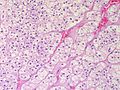
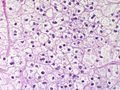
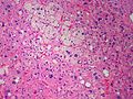
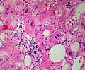
==Microscopic== | |||
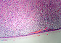
Classic features: | |||
*Well-defined cell borders. | |||
*Clear cells (abundant, finely vacuolated cytoplasm) | |||
*Polygonal pink cells. | |||
*Most of the nuclei are bland, central and round. | |||
*May have foci of [[necrosis]]/degeneration and nuclear atypia. | |||
<gallery> | |||
Image: Adrenal CorticalAdenoma DSCN5001 PA.JPG|Adrenal Cortical Adenoma (SKB) | |||
Image: Adrenal CorticalAdenoma DSCN5002 PA.JPG|Adrenal Cortical Adenoma (SKB) | |||
Image: Adrenal CorticalAdenoma DSCN5004 PA.JPG|Adrenal Cortical Adenoma (SKB) | |||
Image: Adrenal CorticalAdenoma DSCN5005 PA.JPG|Adrenal Cortical Adenoma (SKB) | |||
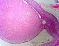
Image: Adrenal CorticalAdenoma MP CTR.jpg|Adrenal Cortical Adenoma - Medium power (SKB) | |||
Image: Adrenal CorticalAdenoma HP CTR.jpg|Adrenal Cortical Adenoma - High power. Abundant clear cytoplasm. Round, regular nuclei. (SKB) | |||
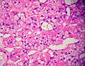
Image: Adrenal CorticalAdenoma MP PA.JPG|Adrenal Cortical Adenoma - Some pleomorphism - Medium power (SKB) | |||
Image: Adrenal LipoAdenoma MP PA.JPG|Adrenal cortical adenoma with fat - "lipoadenoma" (SKB) | |||
</gallery> | |||
Note: | |||
*In aldosterone producing tumours: | |||
**May extend outside of the capsule (should ''not'' be diagnosed as ''[[adrenal cortical carcinoma]]''). | |||
**No atrophy of non-hyperplastic cortex. | |||
**May show spironolactone bodies if hypertension treated with spironolactone prior to surgery. | |||
DDx: | |||
*Adrenal cortical nodule.<ref name=Ref_EP200>{{Ref EP|200}}</ref> | |||
*[[Adrenal cortical hyperplasia]]. | |||
**Hyperplasia is multifocal.<ref>IAV. 18 February 2009.</ref> | |||
*[[Adrenal cortical carcinoma]]. | |||
==See also== | |||
*[[Adrenal gland]]. | |||
==References== | |||
{{Refist|1}} | |||
[[Category:Adrenal gland]] | |||
[[Category:Diagnosis]] | [[Category:Diagnosis]] | ||
Revision as of 05:53, 9 May 2015
Adrenal cortical adenoma, also adrenocortical adenoma and adrenal adenoma, is a relatively common benign pathology of the adrenal gland.
General
Epidemiology:
- Often an incidental finding.
Pathologic/clinical:
- May be hormonally active.
- Can be a cause of hypertension.[1]
- Radiologists are good at identifying adenomas, as they are usually lipid rich and have a characteristic low HU signal.[2]
Indications for excision:[4][5]
- Lesions >30 mm.
- Hormonally active.
- Non-incidental finding. (???)
- Adrenal vein sampling (AVS) suggestive of adenoma.[1]
Notes:
- Cushing disease is due to the ACTH over-production by the pituitary.
- In cortisol producing tumours (Cushing syndrome): atrophy of the non-hyperplastic cortex (due to feedback inhibition from the pituitary gland).
Microscopic
Classic features:
- Well-defined cell borders.
- Clear cells (abundant, finely vacuolated cytoplasm)
- Polygonal pink cells.
- Most of the nuclei are bland, central and round.
- May have foci of necrosis/degeneration and nuclear atypia.
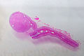
Adrenal Cortical Adenoma (SKB)
Adrenal Cortical Adenoma (SKB)
Adrenal Cortical Adenoma (SKB)
Adrenal Cortical Adenoma (SKB)
Adrenal Cortical Adenoma - Medium power (SKB)
Adrenal Cortical Adenoma - High power. Abundant clear cytoplasm. Round, regular nuclei. (SKB)
Adrenal Cortical Adenoma - Some pleomorphism - Medium power (SKB)
Adrenal cortical adenoma with fat - "lipoadenoma" (SKB)


Note:
- In aldosterone producing tumours:
- May extend outside of the capsule (should not be diagnosed as adrenal cortical carcinoma).
- No atrophy of non-hyperplastic cortex.
- May show spironolactone bodies if hypertension treated with spironolactone prior to surgery.
DDx:
- Adrenal cortical nodule.[6]
- Adrenal cortical hyperplasia.
- Hyperplasia is multifocal.[7]
- Adrenal cortical carcinoma.
See also
References
- ↑ 1.0 1.1 1.2 Myint, KS.; Watts, M.; Appleton, DS.; Lomas, DJ.; Jamieson, N.; Taylor, KP.; Coghill, S.; Brown, MJ. (Jun 2008). "Primary hyperaldosteronism due to adrenal microadenoma: a curable cause of refractory hypertension.". J Renin Angiotensin Aldosterone Syst 9 (2): 103-6. doi:10.3317/jraas.2008.015. PMID 18584586.
- ↑ URL: http://emedicine.medscape.com/article/376240-overview.
- ↑ Fujiwara, M.; Murao, K.; Imachi, H.; Yoshida, K.; Muraoka, T.; Ohyama, T.; Kushida, Y.; Haba, R. et al. (Oct 2010). "Misdiagnosis of two cases of primary aldosteronism owing to failure of computed tomography to detect adrenal microadenoma.". Am J Med Sci 340 (4): 335-7. doi:10.1097/MAJ.0b013e3181e95587. PMID 20881759.
- ↑ Luton, JP.; Martinez, M.; Coste, J.; Bertherat, J. (Jul 2000). "Outcome in patients with adrenal incidentaloma selected for surgery: an analysis of 88 cases investigated in a single clinical center.". Eur J Endocrinol 143 (1): 111-7. PMID 10870039.
- ↑ Liu, XK.; Liu, XJ.; Dong, X.; Kong, CZ. (Jun 2008). "[Clinical research about treatment for adrenal incidentalomas]". Zhonghua Wai Ke Za Zhi 46 (11): 832-4. PMID 19035218.
- ↑ Thompson, Lester D. R. (2006). Endocrine Pathology: A Volume in Foundations in Diagnostic Pathology Series (1st ed.). Churchill Livingstone. pp. 200. ISBN 978-0443066856.
- ↑ IAV. 18 February 2009.