Difference between revisions of "Acute myeloid leukemia"
Jump to navigation
Jump to search
(redirect) |
m (hive off from Leukemia page) |
||
| Line 1: | Line 1: | ||
Abbreviated ''[[AML]]''. | |||
[[Category: | ===General=== | ||
*Adults. | |||
Exclusions for this diagnosis: | |||
*Prior [[MDS]]. | |||
*[[Down syndrome]]. | |||
===Microscopic=== | |||
Features: | |||
*[[Auer rods]] present | |||
*Cytoplasmic granularity. | |||
*Large cells. | |||
Note: | |||
*May be classified by morphology, using the (old) French-American-British (FAB) classification (M0-M7). | |||
====Image==== | |||
<gallery> | |||
Image:Auer_rods.PNG | Auer rods in an AML. (WC) | |||
</gallery> | |||
www: | |||
*[http://path.upmc.edu/cases/case344.html AML - several images (upmc.edu)]. | |||
===Molecular=== | |||
*Must exclude all the recurrent cytogenetic abnormalities - see below. | |||
==AML with recurrent cytogenetic abnormalities== | |||
===Acute myeloid leukemia with t(8;21)=== | |||
*t(8;21)(q22;q22).<ref>{{Cite journal | last1 = Berger | first1 = R. | title = Translocation t(8;21)(q22;q22): cytogenetics and molecular biology. | journal = Nouv Rev Fr Hematol | volume = 36 Suppl 1 | issue = | pages = S67-9 | month = | year = 1994 | doi = | PMID = 8177719 }}</ref> | |||
IHC: | |||
*CD34+, CD13+, MPO+ (cytoplasm), CD33+ (weak). | |||
*[[CD56]]+, CD117+. | |||
**Usu. assoc. with a bad prognosis. | |||
Flow cytometry: | |||
*CD19+, PAX5+, CD79a +/-. | |||
Images: | |||
*[http://path.upmc.edu/cases/case712.html AML with t(8;21) (upmc.edu)]. | |||
===Acute myeloid leukemia with inv(16)=== | |||
*inv(16)(p13.1q22).<ref name=pmid16917916>{{Cite journal | last1 = Lu | first1 = CM. | last2 = Murata-Collins | first2 = JL. | last3 = Wang | first3 = E. | last4 = Siddiqi | first4 = I. | last5 = Lawrence | first5 = HJ. | title = Concurrent acute myeloid leukemia with inv(16)(p13.1q22) and chronic lymphocytic leukemia: molecular evidence of two separate diseases. | journal = Am J Hematol | volume = 81 | issue = 12 | pages = 963-8 | month = Dec | year = 2006 | doi = 10.1002/ajh.20716 | PMID = 16917916 }} | |||
</ref> | |||
*Associated with [[myeloid sarcoma]]. | |||
Microscopic: | |||
*Blast count usu. ~20% (low). | |||
*Eosinophilic granules. | |||
**Used to be classified as "M4" with eosinophilia. | |||
IHC: | |||
*CD2+ -- common. | |||
===Acute myeloid leukemia with t(15;17)=== | |||
*[[AKA]] ''acute promyelocytic leukemia'' | |||
**Abbreviated ''APL''. | |||
*t(15;17)(q22;q12). | |||
**Fusion transcripts: PML<ref name=omim102578>{{OMIM|102578}}</ref>-RARA.<ref name=omim180240>{{OMIM|180240}}</ref> | |||
====General==== | |||
Clinical: | |||
*Associated with [[DIC]]. | |||
*Treatment: all-trans retinoic acid (ATRA). | |||
Variants: | |||
*t(11;17) -- ATRA doesn't work.<ref>{{Ref APBR|623 Q2}}</ref> | |||
*t(17;17) -- ATRA doesn't work. | |||
*t(5;17). (???) | |||
====Microscopic==== | |||
Comes in two flavours. | |||
Microscopic (Hypergranular ''or'' typical APL): | |||
*Bean-shaped nucleus ''or'' bilobed nucleus. | |||
*Buddles of Auer rods - known as "Faggot cells". | |||
Microscopic (Microgranular ''or'' hypogranular APL): | |||
*Bilobed nuclei with nuclear overlap. (???) | |||
*Absence of granules on light microscopy. | |||
=====Images===== | |||
<gallery> | |||
Image:Faggot cell in AML-M3.jpg |Faggot cell in AML-M3. (WC) | |||
</gallery> | |||
www: | |||
*[http://path.upmc.edu/cases/case457/images/fig01.jpg AML - showing Auer rods (upmc.edu)].<ref>URL: [http://path.upmc.edu/cases/case457.html http://path.upmc.edu/cases/case457.html]. Accessed on: 21 January 2012.</ref> | |||
*[http://path.upmc.edu/cases/case705.html APML - several images (upmc.edu)]. | |||
====IHC==== | |||
*CD2 +ve, CD34 +ve/-ve, CD56 +ve/-ve. | |||
====[[Flow cytometry]]==== | |||
*CD34 -ve, HLA-DR -ve. | |||
*CD33 +ve, CD13 +ve/-ve, CD117 +ve (weak), CD56 +ve/-ve. | |||
===Acute myeloid leukemia with t(9;11)=== | |||
*t(9;11). | |||
Microscopic: | |||
*Monoblastic morphology. (???) | |||
*Myelomonocytic morphology. (???) | |||
Clinical: | |||
*+/-[[DIC]]. | |||
*Usu. children. | |||
IHC: | |||
*CD33+, CD65+, CD4+, HLA-DR+. | |||
*CD34+. (???) | |||
*CD13+. (???) | |||
[[Category: Hematopathology]] | |||
Revision as of 18:48, 26 May 2018
Abbreviated AML.
General
- Adults.
Exclusions for this diagnosis:
- Prior MDS.
- Down syndrome.
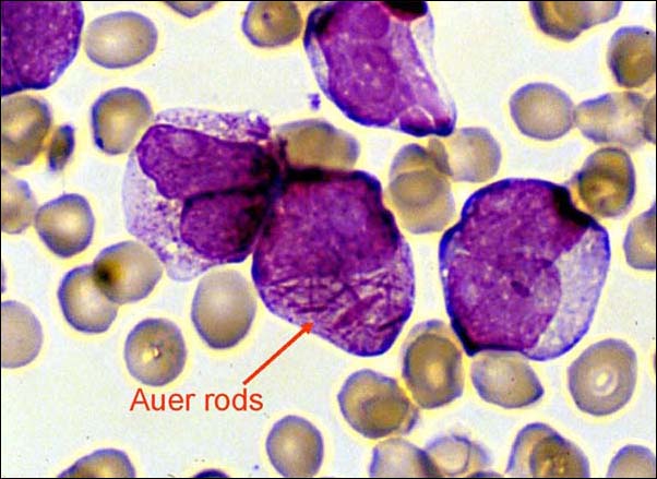
Microscopic
Features:
- Auer rods present
- Cytoplasmic granularity.
- Large cells.
Note:
- May be classified by morphology, using the (old) French-American-British (FAB) classification (M0-M7).
Image
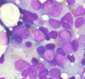
Auer rods in an AML. (WC)

www:
Molecular
- Must exclude all the recurrent cytogenetic abnormalities - see below.
AML with recurrent cytogenetic abnormalities
Acute myeloid leukemia with t(8;21)
- t(8;21)(q22;q22).[1]
IHC:
- CD34+, CD13+, MPO+ (cytoplasm), CD33+ (weak).
- CD56+, CD117+.
- Usu. assoc. with a bad prognosis.
Flow cytometry:
- CD19+, PAX5+, CD79a +/-.
Images:
Acute myeloid leukemia with inv(16)
- inv(16)(p13.1q22).[2]
- Associated with myeloid sarcoma.
Microscopic:
- Blast count usu. ~20% (low).
- Eosinophilic granules.
- Used to be classified as "M4" with eosinophilia.
IHC:
- CD2+ -- common.
Acute myeloid leukemia with t(15;17)
- AKA acute promyelocytic leukemia
- Abbreviated APL.
- t(15;17)(q22;q12).
General
Clinical:
- Associated with DIC.
- Treatment: all-trans retinoic acid (ATRA).
Variants:
- t(11;17) -- ATRA doesn't work.[5]
- t(17;17) -- ATRA doesn't work.
- t(5;17). (???)
Microscopic
Comes in two flavours.
Microscopic (Hypergranular or typical APL):
- Bean-shaped nucleus or bilobed nucleus.
- Buddles of Auer rods - known as "Faggot cells".
Microscopic (Microgranular or hypogranular APL):
- Bilobed nuclei with nuclear overlap. (???)
- Absence of granules on light microscopy.
Images
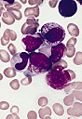
Faggot cell in AML-M3. (WC)

www:
IHC
- CD2 +ve, CD34 +ve/-ve, CD56 +ve/-ve.
Flow cytometry
- CD34 -ve, HLA-DR -ve.
- CD33 +ve, CD13 +ve/-ve, CD117 +ve (weak), CD56 +ve/-ve.
Acute myeloid leukemia with t(9;11)
- t(9;11).
Microscopic:
- Monoblastic morphology. (???)
- Myelomonocytic morphology. (???)
Clinical:
- +/-DIC.
- Usu. children.
IHC:
- CD33+, CD65+, CD4+, HLA-DR+.
- CD34+. (???)
- CD13+. (???)
- ↑ Berger, R. (1994). "Translocation t(8;21)(q22;q22): cytogenetics and molecular biology.". Nouv Rev Fr Hematol 36 Suppl 1: S67-9. PMID 8177719.
- ↑ Lu, CM.; Murata-Collins, JL.; Wang, E.; Siddiqi, I.; Lawrence, HJ. (Dec 2006). "Concurrent acute myeloid leukemia with inv(16)(p13.1q22) and chronic lymphocytic leukemia: molecular evidence of two separate diseases.". Am J Hematol 81 (12): 963-8. doi:10.1002/ajh.20716. PMID 16917916.
- ↑ Online 'Mendelian Inheritance in Man' (OMIM) 102578
- ↑ Online 'Mendelian Inheritance in Man' (OMIM) 180240
- ↑ Lefkowitch, Jay H. (2006). Anatomic Pathology Board Review (1st ed.). Saunders. pp. 623 Q2. ISBN 978-1416025887.
- ↑ URL: http://path.upmc.edu/cases/case457.html. Accessed on: 21 January 2012.