Difference between revisions of "Acute infectious pneumonia"
Jump to navigation
Jump to search
m (chg redirect) |
|||
| (2 intermediate revisions by the same user not shown) | |||
| Line 1: | Line 1: | ||
# | '''Acute infectious pneumonia''' is a common type of [[pneumonia]]. It is usually diagnosed clinically and uncommonly biopsied. | ||
==General== | |||
Clinical features: | |||
*[[Dyspnea]]. | |||
*Chest pain. | |||
*Fever. | |||
It is seen by pathologists at [[autopsy]] from time-to-time, and in advanced [[lung cancer]]. | |||
===Etiology=== | |||
Most common cause: | |||
*''Streptococcus pneumoniae''.<ref name=Ref_PBoD8_711>{{Ref PBoD8|711}}</ref> | |||
The top three community acquired (acute) pneumonia:<ref name=pmid12239229>{{Cite journal | last1 = Nicolau | first1 = D. | title = Clinical and economic implications of antimicrobial resistance for the management of community-acquired respiratory tract infections. | journal = J Antimicrob Chemother | volume = 50 Suppl S1 | issue = | pages = 61-70 | month = Sep | year = 2002 | doi = | PMID = 12239229 }}</ref> | |||
*''Streptococcuc pneumonia''. | |||
*''Haemophilus influenzae''. | |||
*''Moraxella catarrhalis''. | |||
Other community acquired pneumonia:<ref name=Ref_PBoD8_711>{{Ref PBoD8|711}}</ref> | |||
*S. aureus. | |||
*Legionaella pneumophila. | |||
*Klebsiella pneumoniae. | |||
*[[Pseudomonas]]. | |||
Hospital-acquired pneumonia:<ref name=Ref_PBoD8_711>{{Ref PBoD8|711}}</ref> | |||
*Gram-negative rods. | |||
*''Staphylococcus aureus''. | |||
==Radiologic correlate== | |||
*Air space disease. | |||
==Gross pathology== | |||
*Consolidation (the lung parenchyma is firm) - best appreciated by running a finger over the cut surface of the lung with a small-to-moderate amount of pressure. | |||
Bronchopneumonia: | |||
*Classically yellow-white centered on the bronchi.<ref>{{Ref AoGP|93}}</ref> | |||
Lobar pneumnia is classically described in four stages:<ref>{{Ref AoGP|92}}</ref><ref>URL: [http://www.histopathology-india.net/Lobar_Pneumonia.htm http://www.histopathology-india.net/Lobar_Pneumonia.htm]. Accessed on: 27 February 2012.</ref> | |||
#Congestion - day 1-2. | |||
#Red hepatization - day 2-4. | |||
#Gray hepatization - day 4-6. | |||
#Resolution - day 6+. | |||
Note: | |||
*The stages of lobar pneumonia is considered more-or-less historical. In the age of antibiotics, lobar pneumonia is uncommon. | |||
==Microscopic== | |||
Features: | |||
*Alveoli packed with [[PMN]]s. | |||
*+/-Clusters of bacteria - small dots or rods. | |||
*+/-Abscess formation. | |||
**Lung abscess = destruction of parenchyma + [[PMN]]s.<ref name=Ref_AoGP95>{{Ref AoGP|95}}</ref> | |||
DDx: | |||
*[[Aspiration pneumonia]] - aspirated material, usually lack microorganisms. | |||
===Images=== | |||
<gallery> | |||
Image:Pneumonia_alveolus.jpg | Normal alveoli & pneumonia. (WC) | |||
</gallery> | |||
<gallery> | |||
Image: Acute pneumonia -- low mag.jpg | AP - low mag. (WC) | |||
Image: Acute pneumonia -- intermed mag.jpg | AP - intermed. mag. (WC) | |||
Image: Acute pneumonia - alt -- intermed mag.jpg | AP - intermed. mag. (WC) | |||
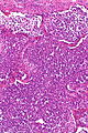
Image: Acute pneumonia -- high mag.jpg | AP - high mag. (WC) | |||
Image: Acute pneumonia -- very high mag.jpg | AP - very high mag. (WC) | |||
</gallery> | |||
==Stains== | |||
*Gram stain -- to type the bacteria. | |||
==See also== | |||
*[[Pneumonia]]. | |||
*[[Acute pneumonia]]. | |||
==References== | |||
{{Reflist|1}} | |||
[[Category:Diagnosis]] | [[Category:Diagnosis]] | ||
[[Category:Medical lung diseases]] | |||
Latest revision as of 16:01, 13 February 2016
Acute infectious pneumonia is a common type of pneumonia. It is usually diagnosed clinically and uncommonly biopsied.
General
Clinical features:
- Dyspnea.
- Chest pain.
- Fever.
It is seen by pathologists at autopsy from time-to-time, and in advanced lung cancer.
Etiology
Most common cause:
- Streptococcus pneumoniae.[1]
The top three community acquired (acute) pneumonia:[2]
- Streptococcuc pneumonia.
- Haemophilus influenzae.
- Moraxella catarrhalis.
Other community acquired pneumonia:[1]
- S. aureus.
- Legionaella pneumophila.
- Klebsiella pneumoniae.
- Pseudomonas.
Hospital-acquired pneumonia:[1]
- Gram-negative rods.
- Staphylococcus aureus.
Radiologic correlate
- Air space disease.
Gross pathology
- Consolidation (the lung parenchyma is firm) - best appreciated by running a finger over the cut surface of the lung with a small-to-moderate amount of pressure.
Bronchopneumonia:
- Classically yellow-white centered on the bronchi.[3]
Lobar pneumnia is classically described in four stages:[4][5]
- Congestion - day 1-2.
- Red hepatization - day 2-4.
- Gray hepatization - day 4-6.
- Resolution - day 6+.
Note:
- The stages of lobar pneumonia is considered more-or-less historical. In the age of antibiotics, lobar pneumonia is uncommon.
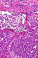
Microscopic
Features:
- Alveoli packed with PMNs.
- +/-Clusters of bacteria - small dots or rods.
- +/-Abscess formation.
DDx:
- Aspiration pneumonia - aspirated material, usually lack microorganisms.
Images
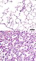
Normal alveoli & pneumonia. (WC)

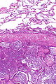
AP - low mag. (WC)
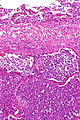
AP - intermed. mag. (WC)
AP - intermed. mag. (WC)
AP - high mag. (WC)
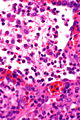
AP - very high mag. (WC)


Stains
- Gram stain -- to type the bacteria.
See also
References
- ↑ 1.0 1.1 1.2 Kumar, Vinay; Abbas, Abul K.; Fausto, Nelson; Aster, Jon (2009). Robbins and Cotran pathologic basis of disease (8th ed.). Elsevier Saunders. pp. 711. ISBN 978-1416031215.
- ↑ Nicolau, D. (Sep 2002). "Clinical and economic implications of antimicrobial resistance for the management of community-acquired respiratory tract infections.". J Antimicrob Chemother 50 Suppl S1: 61-70. PMID 12239229.
- ↑ Rose, Alan G. (2008). Atlas of Gross Pathology with Histologic Correlation (1st ed.). Cambridge University Press. pp. 93. ISBN 978-0521868792.
- ↑ Rose, Alan G. (2008). Atlas of Gross Pathology with Histologic Correlation (1st ed.). Cambridge University Press. pp. 92. ISBN 978-0521868792.
- ↑ URL: http://www.histopathology-india.net/Lobar_Pneumonia.htm. Accessed on: 27 February 2012.
- ↑ Rose, Alan G. (2008). Atlas of Gross Pathology with Histologic Correlation (1st ed.). Cambridge University Press. pp. 95. ISBN 978-0521868792.