Metaphyseal fibrous defect
Jump to navigation
Jump to search
| Metaphyseal fibrous defect | |
|---|---|
| Diagnosis in short | |
|
| |
| Synonyms | Nonossifying fibroma |
| Clinical history | Incidental radiograhic finding |
| Radiology | Lucent defect |
Metaphyseal fibrous defect, abbreviated MFD, is a common benign abnormality of the metaphysis, classically seen in children and young adults.
They are also known as fibrous cortical defect, fibrous metaphyseal defect, and fibroxanthoma of bone. Nonossifying fibroma is a larger lesion but otherwise identical.
General
- Common.
- Non-neoplastic.
- Self-limited.
- Skeletally immature individuals, children and adolescents.
- Often small lesions discovered as an radiographic incidentaloma.
- Rarely seen as a pathologic specimen (should not be biopsied).
- May be seen in the context of Jaffe-Campanacci syndrome.[1]
Clinical history:
- Incidental radiographic finding.
- Pathologic fracture.
Prognosis:
- Ideally should not be biopsied.
- Radiographically characteristic and benign.
- Ideally should not be treated or even biopsied.
- Spontaneously resolve by ossification.
- May resolve into a 'bone island'.
Site
- Metaphysis of distal femur or proximal tibia (80%).
- Cortical.
- Metaphysis.
- Long bones.
- Eccentric location.
Gross
- Firm, granular, brown to yellow to red.
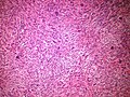
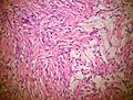
Microscopic
Features:
- Spindle cells without cytologic atypia are arranged in a storiform pattern.
- Scattered chronic inflammatory cells and benign giant cells.
- Foam cells and hemosiderin deposition are present.
- Mitoses are seen but cytologic atypia is absent.
DDx (microscopic):
- Giant cell tumour of bone - epiphyseal location, occurs in adults.
Relevant Diagnostic Groups
- Clinical
- Pathologic
- Giant cell lesions of bone.
- Spindle cell lesions of bone.
Images
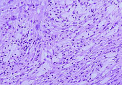
Metaphyseal fibrous defect - intermediate magnification.]]
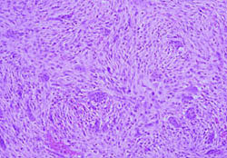
Metaphyseal fibrous defect - high magnification.]]


www:
%20micro.jpg)
Stains
- Not relevant.
IHC
- Not relevant
Molecular
- Not relevant
Radiographic findings
Sharply demarcated, lucent, loculated, meta-diaphyseal lesion surrounded by a rim of sclerotic bone
Sign out
BONE, CURETTAGE: METAPHYSEAL FIBROUS DEFECT / NONOSSIFYING FIBROMA.
See also
- Bone.
References
- ↑ URL: http://www.bonetumor.org/plasma-cell-tumors/jaffe-campanacci-syndrome. Accessed on: October 14, 2014.
- ↑ URL: http://radiopaedia.org/articles/lytic-bone-lesion-mnemonic. Accessed on: October 14, 2014.
- ↑ URL: http://radiopaedia.org/articles/skeletal-do-not-touch-lesions-1. Accessed on: October 14, 2014.