Uterine adenomyosis
Jump to navigation
Jump to search
Uterine adenomyosis, also adenomyosis of the uterus, is a relatively common benign pathology of the uterine corpus. It can be thought of as endometriosis in the uterine smooth muscle.
| Uterine adenomyosis | |
|---|---|
| Diagnosis in short | |
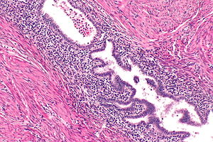
 Uterine adenomyosis. H&E stain. | |
|
| |
| LM | at least 2 of 3: (1) endometrial glands, (2) endometrial stroma, (3) hemosiderin-laden macrophages |
| LM DDx | endometrioid endometrial carcinoma |
| Gross | globoid shape, slightly enlarged, trabeculated cut surface +/- small foci of hemorrhage |
| Site | uterus |
|
| |
| Associated Dx | endometriosis (?) |
| Signs | menorrhagia |
| Symptoms | cyclic pelvic pain, dysmenorrhea |
| Prevalence | common |
| Prognosis | benign |
Uterine adenomyoma redirects here.
General
- Common.
- May be a cause of bleeding.[1]
- Dysmenorrhea - painful menses.[2]
- Associated with endometriosis.[citation needed]
Gross
Features:
- Trabeculated cut surface +/- small foci of hemorrhage.[3]
- Often described as "basket-weave" pattern.
- Globoid, slightly enlarged.[4]
Note:
- May form a mass - known as adenomyoma.[5]
Image:
Microscopic
Features:
- Endometrial glands within uterine muscle - key feature.
- Endometrial glands:
- Circular.
- Simple epithelial or pseudostratified epithelium +/- mitoses.
- +/-Surrounded by endometrial stroma.
- Densely packed spindle cells without nuclear atypia.
- Blood:
- Within glands.
- Hemosiderin-laden macrophages.
- Endometrial glands:
Note:
- Can be thought of as endometriosis of the myometrium.
- A large number of extent criteria exist - see extent criteria.[6]
DDx:
Extent criteria
Consensus is lacking on diagnostic criteria. Focal superficial endometrial glands are generally not deemed sufficient by most.[6]
Images
Sign out
Uterus, Bilateral Fallopian Tubes, Hysterectomy and Bilateral Salpingectomy:
- Uterine adenomyosis, extensive.
- Uterine leiomyoma.
- Proliferative endometrium.
- Fallopian tubes within normal limits.
- NEGATIVE for malignancy.
Block letters
UTERUS, UTERINE CERVIX, TOTAL HYSTERECTOMY:
- UTERUS WITH ADENOMYOSIS.
- UTERINE CERVIX WITHIN NORMAL LIMITS.
- PROLIFERATIVE PHASE ENDOMETRIUM.
UTERUS, UTERINE CERVIX, TOTAL HYSTERECTOMY:
- UTERUS WITH SUPERFICIAL ADENOMYOSIS.
- UTERINE CERVIX WITH PARTIAL DENUDATION, FOCUS OF ENDOMETRIOSIS AND INFLAMMATION,
  OTHERWISE WITHIN NORMAL LIMITS.
- SUPERFICIAL FIBROSIS AND HYALINE CHANGE OF THE UTERINE LINING -- COMPATIBLE
  WITH PRIOR ABLATION.
See also
References
- ↑ Reinhold, C.; Tafazoli, F.; Mehio, A.; Wang, L.; Atri, M.; Siegelman, ES.; Rohoman, L. (Oct 1999). "Uterine adenomyosis: endovaginal US and MR imaging features with histopathologic correlation.". Radiographics 19 Spec No: S147-60. PMID 10517451.
- ↑ Cockerham, AZ.. "Adenomyosis: a challenge in clinical gynecology.". J Midwifery Womens Health 57 (3): 212-20. doi:10.1111/j.1542-2011.2011.00117.x. PMID 22594861.
- ↑ Lester, Susan Carole (2010). Manual of Surgical Pathology (3rd ed.). Saunders. pp. 432. ISBN 978-0-323-06516-0.
- ↑ HUNTER, WC.; SMITH, LL.; REINER, WC. (Apr 1947). "Uterine adenomyosis; incidence, symptoms, and pathology in 1,856 hysterectomies.". Am J Obstet Gynecol 53 (4): 663-8. PMID 20291238.
- ↑ Tahlan, A.; Nanda, A.; Mohan, H. (Oct 2006). "Uterine adenomyoma: a clinicopathologic review of 26 cases and a review of the literature.". Int J Gynecol Pathol 25 (4): 361-5. doi:10.1097/01.pgp.0000209570.08716.b3. PMID 16990713.
- ↑ 6.0 6.1 Abbott JA (April 2017). "Adenomyosis and Abnormal Uterine Bleeding (AUB-A)-Pathogenesis, diagnosis, and management". Best Pract Res Clin Obstet Gynaecol 40: 68–81. doi:10.1016/j.bpobgyn.2016.09.006. PMID 27810281.