Craniopharyngioma
Revision as of 11:39, 20 March 2015 by Jensflorian (talk | contribs) (→General: mutation profile added)
Craniopharyngioma is a benign neuropathology tumour.
| Adamantinomatous craniopharyngioma | |
|---|---|
| Diagnosis in short | |
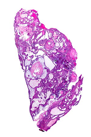
 Adamantinomatous craniopharyngioma. HPS stain. | |
|
| |
| LM | well-circumscribed (or pseudoinvasive border), multicystic, small-to-medium sized cells with moderate amount of basophilic cytoplasm, bland nuclei (with occ. small nucleoli), "wet" keratin (nests of whorled keratin), calcifications |
| Gross | cystic mass filled with motor oil-like fluid |
| Site | sella turcica |
|
| |
| Clinical history | adults & children |
| Radiology | classically calcified |
| Prognosis | benign |
| Clin. DDx | other sella turcica lesions |
| Papillary craniopharyngioma | |
|---|---|
| Diagnosis in short | |
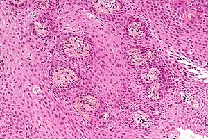
 Papillary craniopharyngioma. HPS stain. | |
|
| |
| LM | non-keratinized squamous epithelium (without nuclear atypia), fibrovascular cores (required for papillary) |
| Site | sella turcica |
|
| |
| Clinical history | adults |
| Prognosis | benign |
| Clin. DDx | other sella turcica lesions |
It is subdivided into papillary craniopharyngioma and adamantinomatous craniopharyngioma.
General
- Develop from remains of Rathke's pouch or squamous epithelial cell rests.[1]
Subtypes:[1]
- Adamantinomatous type.
- Squamous papillary type.
Adamantinomatous
- Adults and children.
- Typically contain mutations in CTNNB1 (the gene that encodes β-catenin).[2]
- Usually intranuclear β-catenin immunohistochemical positivity.
Papillary
Gross
- Cystic mass filled with motor oil-like fluid.[5]
- May not be seen in the papillary variant of craniopharyngioma.
Radiology:[1]
- Calcified - adamantinomatous type only.
- Solid & cystic.
Microscopic
Adamantinomatous
Features (adamantinomatous):[6]
- Well-circumscribed (or pseudoinvasive border).
- Multicystic.
- Small-to-medium sized cells with moderate amount of basophilic cytoplasm.
- Bland nuclei (with occ. small nucleoli).
- "Wet" keratin - nests of whorled keratin.
- Calcifications (non-psammomatous).
Images
Papillary
Features (papillary):[7]
- Non-keratinized squamous epithelium (without nuclear atypia).
- Fibrovascular cores (required for papillary).
Notes:
- +/-Cilia (rare).
- +/-Goblet cell-like formations (rare).
Image
www:
See also
References
- ↑ 1.0 1.1 1.2 Garnett, MR.; Puget, S.; Grill, J.; Sainte-Rose, C. (2007). "Craniopharyngioma.". Orphanet J Rare Dis 2: 18. doi:10.1186/1750-1172-2-18. PMID 17425791.
- ↑ Preda, V.; Larkin, SJ.; Karavitaki, N.; Ansorge, O.; Grossman, AB. (Oct 2014). "The Wnt Signalling Cascade and the Adherens Junction Complex in Craniopharyngioma Tumorigenesis.". Endocr Pathol. doi:10.1007/s12022-014-9341-8. PMID 25355426.
- ↑ Giangaspero, F.; Burger, PC.; Osborne, DR.; Stein, RB. (Jan 1984). "Suprasellar papillary squamous epithelioma ("papillary craniopharyngioma").". Am J Surg Pathol 8 (1): 57-64. PMID 6696166.
- ↑ Brastianos et al. Nat. Genet 2014 doi:10.1038/ng.2868
- ↑ Fernandez-Miranda, JC.; Gardner, PA.; Snyderman, CH.; Devaney, KO.; Strojan, P.; Suárez, C.; Genden, EM.; Rinaldo, A. et al. (Jul 2012). "Craniopharyngioma: a pathologic, clinical, and surgical review.". Head Neck 34 (7): 1036-44. doi:10.1002/hed.21771. PMID 21584897.
- ↑ Tadrous, Paul.J. Diagnostic Criteria Handbook in Histopathology: A Surgical Pathology Vade Mecum (1st ed.). Wiley. pp. 184. ISBN 978-0470519035.
- ↑ Perry, Arie; Brat, Daniel J. (2010). Practical Surgical Neuropathology: A Diagnostic Approach: A Volume in the Pattern Recognition series (1st ed.). Churchill Livingstone. pp. 406. ISBN 978-0443069826.
- ↑ URL: http://library.med.utah.edu/WebPath/jpeg4/ENDO115.jpg. Accessed on: 6 December 2010.