Carcinoma ex pleomorphic adenoma
Carcinoma ex pleomorphic adenoma, abbreviated ca ex PA, is a rare malignant salivary gland tumour that arises from a pleomorphic adenoma.
| Carcinoma ex pleomorphic adenoma | |
|---|---|
| Diagnosis in short | |
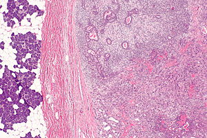
 Carcinoma ex pleomorphic adenoma. H&E stain. | |
|
| |
| LM | epithelial or myoepithelial cells with cytologic features of malignancy, pleomorphic adenoma (required if no history) |
| LM DDx | salivary duct carcinoma, mucoepidermoid carcinoma, adenoid cystic carcinoma, pleomorphic adenoma |
| Gross | firm |
| Site | salivary gland - usu. parotid gland |
|
| |
| Clinical history | pleomorphic adenoma (required if not present in assoc. with carcinoma) |
| Signs | usu. mass lesion, firm |
| Prevalence | uncommon |
| Prognosis | dependent on stage, typically poor |
General
Definition:
- Malignancy arising from a pleomorphic adenoma.[1]
Diagnosis (either 1 or 2):
- History of a pleomorphic adenoma at the same site (~25% of cases[1]).
- Features of a pleomorphic adenoma and a carcinoma.
Epidemiology:
Gross
- Usually mass lesion - typically parotid gland.[1]
Microscopic
Features:
- Cells (epithelial or myoepithelial) with cytologic features of malignancy - key feature.
- Architecture (any of the following):
- Glands.
- Nests.
- Single cells (may be subtle).
Subtyping/patterns:
- Epithelial carcinoma (~75% of cases[1]):
- Ductal carcinoma NOS (arising from ductal cells) - most common pattern for ca ex PA.
- "Named carcinoma":
- Salivary duct carcinoma - second most common pattern for Ca ex PA.
- Mucoepidermoid carcinoma.
- Adenoid cystic carcinoma.
- Myoepithelial carcinoma NOS (arising from myoepithelial cells).
Notes:
- Myoepithelial cells may be clear cells.
DDx:
Subclassification
Extent of invasion:[3]
- Non-invasive AKA intracapsular AKA in situ.
- Minimally invasive <=1.5 mm beyond the capsule.
- Widely invasive >1.5 mm beyond the capsule.
Images
www:
IHC
Features - ca ex PA versus PA:[2]
- Ki-67 >35% of cells.
- HER2 +ve membrane staining (3+ for high-grade, 2+ or less for low-grade).
- p53 +ve.
- AR +ve.
Others:[2]
- CK7 +ve.
- AE1/AE3 +ve.
Sign out
PAROTID GLAND (MASS), LEFT, EXCISION:
- CARCINOMA EX PLEOMORPHIC ADENOMA, LOW-GRADE, INTRACAPSULAR.
-- MARGINS NEGATIVE.
-- SEE TUMOUR SUMMARY AND COMMENTS.
- THREE BENIGN LYMPH NODES.
Micro
The sections show a lesion with spindled (myoepithelial) cells and an epithelial component, on a background of a chondromyxoid stroma. The lesion is encapsulated by a thin layer of fibrous tissue.
Focally, within the lesion, nuclear atypia and mitotic activity are identified, including an atypical mitotic figure. The atypical region stains with p53. Approximately 45% of the atypical cells stain with Ki-67. There is no appreciable HER2 staining.
Unremarkable parotid gland and lymph nodes are present.
See also
References
- ↑ 1.0 1.1 1.2 1.3 1.4 1.5 Antony, J.; Gopalan, V.; Smith, RA.; Lam, AK. (Mar 2012). "Carcinoma ex pleomorphic adenoma: a comprehensive review of clinical, pathological and molecular data.". Head Neck Pathol 6 (1): 1-9. doi:10.1007/s12105-011-0281-z. PMID 21744105.
- ↑ 2.0 2.1 2.2 Di Palma, S. (Jul 2013). "Carcinoma ex pleomorphic adenoma, with particular emphasis on early lesions.". Head Neck Pathol 7 Suppl 1: S68-76. doi:10.1007/s12105-013-0454-z. PMID 23821206.
- ↑ URL: http://www.cap.org/apps/docs/committees/cancer/cancer_protocols/2011/MajorSalGlands_11protocol.pdf. Accessed on: 2 April 2012.