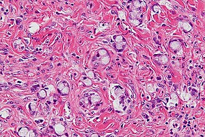
Goblet cell adenocarcinoma
Jump to navigation
Jump to search
Crypt cell carcinoma, also known as goblet cell carcinoid[1][2] and neuroendocrine tumour with goblet cell differentiation, is a rare malignant tumour that is typically seen in the vermiform appendix.
| Goblet cell adenocarcinoma | |
|---|---|
| Diagnosis in short | |
 Crypt cell carcinoma. H&E stain. | |
|
| |
| LM | small clusters of cells with stippled chromatin and a goblet cell-like appearance |
| LM DDx | signet ring cell carcinoma, appendiceal neuroendocrine tumour |
| Stains | alcian blue +ve, PASD +ve, mucicarmine +ve |
| IHC | synaptophysin +ve, chromogranin +ve, S-100 +ve, CK20 +ve |
| Gross | usu. no mass apparent |
| Site | vermiform appendix, elsewhere in the GI tract |
|
| |
| Prevalence | rare |
| Prognosis | moderate |
| Clin. DDx | acute appendicitis, other appendiceal tumours, other abdominal pathology |
General
- Rare appendiceal tumour that typically has an aggressive course vis-a-vis other appendiceal carcinoids.[1]
- Mixed (biphasic) tumour with endocrine and exocrine features.
- Usually presents as acute appendicitis.[3]
- Less common presentations: appendiceal mass, pain.
- Five year survival in one series: 60-85%.[3]
Gross
- Typically no mass is apparent at gross.[3]
Microscopic
Features:[3]
- Mixed neuroendocrine-nonneuroendocrine tumour;[4] features of both carcinoid and adenocarcinoma.[3]
- Archictecture: cells arranged in nests or clusters without a lumen.
- Location: deep to the intestinal crypts (crypts of Lieberkühn); usually do not involve the mucosa.
- Cytoplasm distended with mucin.
- DNA: crescentic nucleus (similar to in signet-ring cells).
- +/-Multinucleation.
- +/-High mitotic rate.
- Usually minimal nuclear atypia.
DDx:
- Appendiceal neuroendocrine tumour.
- Signet ring cell carcinoma - cell detached.
Images
Stains
- Mucin stains +ve:
- Mucicarmine, perodic acid-Schiff diastase (PAS-D), alcian blue.
IHC
- Classic neuroendocrine markers:
- Synaptophysin +ve.
- Chromogranin +ve.
- S-100 +ve.
- NSE +ve.
- Serotonin +ve.
Keratins:
- Usually CK20 +ve > CK7 +ve.
- CEA +ve (membrane).
Notes:
- Nice review of stains in Pahlavan and Kanthan.[3]
See also
References
- ↑ 1.0 1.1 van Eeden S, Offerhaus GJ, Hart AA, et al. (December 2007). "Goblet cell carcinoid of the appendix: a specific type of carcinoma". Histopathology 51 (6): 763–73. doi:10.1111/j.1365-2559.2007.02883.x. PMID 18042066.
- ↑ Pahlavan, PS.; Kanthan, R. (Jun 2005). "Goblet cell carcinoid of the appendix.". World J Surg Oncol 3: 36. doi:10.1186/1477-7819-3-36. PMID 15967038.
- ↑ 3.0 3.1 3.2 3.3 3.4 3.5 Pahlavan PS, Kanthan R (June 2005). "Goblet cell carcinoid of the appendix". World J Surg Oncol 3: 36. doi:10.1186/1477-7819-3-36. PMC 1182398. PMID 15967038. http://wjso.com/content/3/1/36.
- ↑ Volante M, Righi L, Asioli S, Bussolati G, Papotti M (August 2007). "Goblet cell carcinoids and other mixed neuroendocrine/nonneuroendocrine neoplasms". Virchows Arch. 451 Suppl 1: S61–9. doi:10.1007/s00428-007-0447-y. PMID 17684764.