Autosomal dominant polycystic kidney disease
Jump to navigation
Jump to search
| Autosomal dominant polycystic kidney disease | |
|---|---|
| Diagnosis in short | |
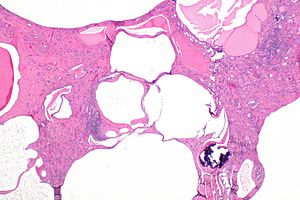
 Micrograph showing polycystic kidney disease in the context of autosomal dominant polycystic kidney disease. H&E stain. | |
|
| |
| LM | cortical cysts lined by simple flattened epithelium, normal renal tubules interspersed between cysts or tubules as seen in end-stage kidney (thyroidization), +/-fibrosis (late-stage), +/-calcifications |
| Subtypes | PKD1 gene associated, PKD2 gene associated |
| LM DDx | acquired cystic renal disease |
| Molecular | mutation in PKD1 gene or PKD2 gene |
| Gross | enlarged kidney composed to many cysts |
| Site | kidney - see cystic kidney diseases |
|
| |
| Associated Dx | end-stage kidney, liver cysts; PKD1 associated: cerebral aneurysms; PKD2 associated: colonic diverticula, aortic aneurysm, mitral valve prolapse |
| Clinical history | family member with polycystic kidney disease |
| Prevalence | uncommon |
| Radiology | polycystic kidneys |
| Prognosis | progressive renal failure |
| Treatment | +/-nephrectomy, dialysis, renal transplant |
Autosomal dominant polycystic kidney disease, abbreviated ADPKD, is a common genetic cause of chronic renal failure.
Surgically removed to due to symptoms (mass effect); native nephrectomy often done concurrently with renal transplant.[1]
General
Etiology
- Mutation in PKD1 gene or PKD2 gene.
- Is classified in a large group of diseases - ciliopathies.
PKD1 related disease:[2]
- Encodes polycystin.
- Death at ~53 years.
- Associated with cerebral aneurysms.
PKD2 related disease:[2]
- Death at ~69 years.
- Associated with colonic diverticula, aortic aneurysm, mitral valve prolapse.
Liver cysts and PKD
General
Features:
- Most common extra-renal manifestation of PKD; dependent on age, sex and renal function:[3]
- Age dependence:
- 10-17% <40 years old have liver cysts.
- 70-75% >60 years old have liver cysts.
- Renal function:
- 60-70% of patients with end-stage renal disease (ESRD) and near-ESRD.
- Females more often affected.
- Age dependence:
- Hepatic function usu. preserved.
Complications:[2]
- Infected cyst.
- Cholangiocarcinoma.
Microscopic
Features:
- Von Meyenburg complexes:
- Cluster of dilated ducts with "altered" bile.
- Surrounded by collagenous stroma.
See: Medical liver disease.
Gross
Features:
- Thin walled cysts.
- Number of cysts:
- If you can count 'em it favours acquired renal cystic disease... if you can't it favours the genetic condition.
- Number of cysts:
Microscopic
Features:[4]
- Cortical cysts lined by simple flattened epithelium.
- Normal renal tubules interspersed between cysts or tubules as seen in end-stage kidney (thyroidization).
- +/-Fibrosis (late-stage).
- +/-Calcifications.[citation needed]
DDx:
- Acquired renal cystic disease - rarely.[5]
Sign out
Left Kidney, Nephrectomy:
- Polycystic kidney with changes of chronic renal failure (thyroidization),
  consistent with polycystic kidney disease.
- NEGATIVE for malignancy.
See also
References
- ↑ Veroux, M.; Zerbo, D.; Basile, G.; Gozzo, C.; Sinagra, N.; Giaquinta, A.; Sanfiorenzo, A.; Veroux, P. (2016). "Simultaneous Native Nephrectomy and Kidney Transplantation in Patients With Autosomal Dominant Polycystic Kidney Disease.". PLoS One 11 (6): e0155481. doi:10.1371/journal.pone.0155481. PMID 27257690.
- ↑ 2.0 2.1 2.2 Burt, Alastair D.;Portmann, Bernard C.;Ferrell, Linda D. (2006). MacSween's Pathology of the Liver (5th ed.). Churchill Livingstone. pp. 174-5. ISBN 978-0-443-10012-3.
- ↑ Perrone RD (June 1997). "Extrarenal manifestations of ADPKD". Kidney Int. 51 (6): 2022–36. PMID 9186898. http://www.nature.com/ki/journal/v51/n6/pdf/ki1997276a.pdf.
- ↑ Fogo, Agnes B.; Kashgarian, Michael (2005). Diagnostic Atlas of Renal Pathology: A Companion to Brenner and Rector's The Kidney 7E (1st ed.). Saunders. pp. 426. ISBN 978-1416028710.
- ↑ Kessler M, Testevuide P, Aymard B, Huu TC (1991). "Acquired renal cystic disease mimicking adult polycystic kidney disease in a patient undergoing long-term hemodialysis". Am. J. Nephrol. 11 (6): 513–7. PMID 1819219.
- ↑ 6.0 6.1 RJ. 20 October 2010.
- ↑ Barbaric, Zoran L. (1994). Principles of Genitourinary Radiology (2nd ed.). Thieme. pp. 87. ISBN 978-0865774933.