Virus
This article collects all things virus. The more general topic of infective thingies is dealt with in microorganisms.
Many virus afflict humans. Only a few of them can be diagnosed histologically.
Viral inclusions - types
Cowdry types:[1]
- Cowdry type A inclusion:[2]
- Round eosinophilic material surrounded by a clear halo.
- Cowdry type B inclusion:[3]
- Neuropathology thingy. (???)
Images:
Viruses
Herpes simplex virus (HSV)
- Canker sores - usually HSV-1.
- Genital herpes - usually HSV-2.
Histology/cytology
Features:[4]
- Clear "ground glass" nuclei.
- Rim of peripheral chromatin.
- Nuclear inclusions.
- Multinucleation with nuclear molding, i.e. multiple nuclei that touch over a large surface area.
Mnemonic - 3 Ms: Margination, Multinucleation, Molding.
Images:
- Herpes simplex virus - multinucleation (virology.org).
- HSV on a Pap test - showing multinucleation (WC).
- HSV esophagitis - very high mag. (WC).
- HSV esophagitis - intermed. mag. (WC).
Cytomegalovirus (CMV)
Microscopic
Features:
- Very large nucleus (as the name implies) with clearing.
- Granular cytoplasmic inclusions (red on H&E sections).
Notes:
- Classically in endothelial cells.
- In the context of esophageal ulcers, it is therefore useful to biopsy the base of the ulcer - if this is suspected.
Images:
Human papilloma virus
- Abbreviated HPV.
Microscopic
Features:
- Koilocytes:
- Perinuclear clearing.
- Nuclear changes.
- Size similar (or larger) to those in the basal layer of the epithelium.
- Nuclear enlargement should be evident on low power, i.e. 25x. [7]
- Central location - nucleus should be smack in the middle of the cell.
Images:
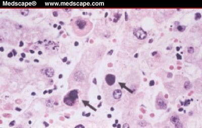
Adenovirus
General
- Common in kids.
- May be seen in the context of (adenovirus) appendicitis.
Microscopic
Features:
- "Smudge" cells[5] - black/blue blob ~ 10-15 micrometers. (???)
Notes:
- May be morphologically similar to CMV, HSV, VZV inclusions.
Images:
- Adenovirus (medscape.com).[6]
- Smudge cell (medpedia.com).
- Necrosis in germinal centre - low mag. (flickr.com).
- Viral inclusions - high mag. (flickr.com).
- IHC for adenovirus (flickr.com)
Parvo B19
Features:
- Big red nuclear inclusion.[7]
See also
References
- ↑ URL: http://www.pathconsultddx.com/pathCon/largeImage?pii=S1559-8675%2806%2970864-6&figureId=fig3&ecomponentId=mmc3. Accessed: 12 January 2010.
- ↑ URL: http://www.whonamedit.com/synd.cfm/3495.html. Accessed on: 22 January 2010.
- ↑ http://www.whonamedit.com/synd.cfm/3496.html. Accessed on: 22 January 2010.
- ↑ SM. 11 January 2010.
- ↑ URL: http://www.pathguy.com/lectures/infect.htm. Accessed on: 8 July 2010.
- ↑ URL:http://www.medscape.com/viewarticle/438534_2. Accessed on: 8 July 2010.
- ↑ URL: http://www.pathguy.com/lectures/infect.htm. Accessed on: 8 July 2010.