Difference between revisions of "Complex sclerosing lesion"
Jump to navigation
Jump to search
| Line 39: | Line 39: | ||
*Associated with subsequent elevated risk of breast cancer.<ref>URL: [http://www.cancer.org/docroot/NWS/content/NWS_1_1x_Radial_Scars.asp http://www.cancer.org/docroot/NWS/content/NWS_1_1x_Radial_Scars.asp]. Accessed on: 4 May 2010.</ref> | *Associated with subsequent elevated risk of breast cancer.<ref>URL: [http://www.cancer.org/docroot/NWS/content/NWS_1_1x_Radial_Scars.asp http://www.cancer.org/docroot/NWS/content/NWS_1_1x_Radial_Scars.asp]. Accessed on: 4 May 2010.</ref> | ||
*Management - usually surgical excision.<ref name=pmid14514771>{{cite journal |author=Kennedy M, Masterson AV, Kerin M, Flanagan F |title=Pathology and clinical relevance of radial scars: a review |journal=J. Clin. Pathol. |volume=56 |issue=10 |pages=721–4 |year=2003 |month=October |pmid=14514771 |pmc=1770086 |doi= |url=}}</ref> | *Management - usually surgical excision.<ref name=pmid14514771>{{cite journal |author=Kennedy M, Masterson AV, Kerin M, Flanagan F |title=Pathology and clinical relevance of radial scars: a review |journal=J. Clin. Pathol. |volume=56 |issue=10 |pages=721–4 |year=2003 |month=October |pmid=14514771 |pmc=1770086 |doi= |url=}}</ref> | ||
**Risk of malignancy for biopsy diagnosed CSLs | **Risk of malignancy on surgical excision for biopsy diagnosed CSLs very small ~1%.<ref name=pmid26772402>{{Cite journal | last1 = Li | first1 = Z. | last2 = Ranade | first2 = A. | last3 = Zhao | first3 = C. | title = Pathologic findings of follow-up surgical excision for radial scar on breast core needle biopsy. | journal = Hum Pathol | volume = 48 | issue = | pages = 76-80 | month = Feb | year = 2016 | doi = 10.1016/j.humpath.2015.06.028 | PMID = 26772402 }}</ref> | ||
*Rare - less than 0.1% of core needle biopsies.<ref name=pmid25578683>{{Cite journal | last1 = Nassar | first1 = A. | last2 = Conners | first2 = AL. | last3 = Celik | first3 = B. | last4 = Jenkins | first4 = SM. | last5 = Smith | first5 = CY. | last6 = Hieken | first6 = TJ. | title = Radial scar/complex sclerosing lesions: a clinicopathologic correlation study from a single institution. | journal = Ann Diagn Pathol | volume = 19 | issue = 1 | pages = 24-8 | month = Feb | year = 2015 | doi = 10.1016/j.anndiagpath.2014.12.003 | PMID = 25578683 }}</ref> | *Rare - less than 0.1% of core needle biopsies.<ref name=pmid25578683>{{Cite journal | last1 = Nassar | first1 = A. | last2 = Conners | first2 = AL. | last3 = Celik | first3 = B. | last4 = Jenkins | first4 = SM. | last5 = Smith | first5 = CY. | last6 = Hieken | first6 = TJ. | title = Radial scar/complex sclerosing lesions: a clinicopathologic correlation study from a single institution. | journal = Ann Diagn Pathol | volume = 19 | issue = 1 | pages = 24-8 | month = Feb | year = 2015 | doi = 10.1016/j.anndiagpath.2014.12.003 | PMID = 25578683 }}</ref> | ||
Revision as of 18:20, 1 January 2017
| Complex sclerosing lesion | |
|---|---|
| Diagnosis in short | |
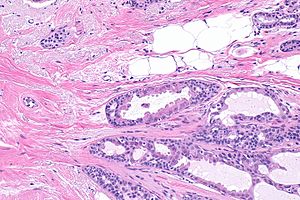
 Complex sclerosing lesion of breast. H&E stain. (WC) | |
|
| |
| Synonyms | radial scar |
|
| |
| LM | stellate lesion (low magnification), center of lesion has fibroelastosis (stroma light pink on H&E), scar like stroma with entrapped normal breast ducts and lobules - glands appear to enlarge with distance from center of lesion |
| LM DDx | invasive ductal carcinoma |
| IHC | myoepithelial cells preset (p63 +ve, calponin +ve) |
| Gross | spiculated mass |
| Site | breast |
|
| |
| Prognosis | benign, increased risk of malignancy |
| Clin. DDx | breast cancer |
| Treatment | excision |
Complex sclerosing lesion (abbreviated CSL), also radial scar, is a benign lesion of the breast that is associated with an increased risk of subsequent breast cancer.
General
- The term radial scar is a misnomer. It isn't a scar. It isn't associated with prior trauma or surgery.[1]
- May appear malignant on imaging.[2]
- Associated with subsequent elevated risk of breast cancer.[4]
- Management - usually surgical excision.[5]
- Risk of malignancy on surgical excision for biopsy diagnosed CSLs very small ~1%.[6]
- Rare - less than 0.1% of core needle biopsies.[7]
Gross
- Spiculated mass.
- Usually small - 3-7 mm.
Image
Microscopic
- Stellate appearance (low magnification).
- Center of lesion has "fibroelastosis" - stroma light pink (on H&E) - key feature.
- Scar like stroma with entrapped normal breast ducts and lobules.
- Glands appear to enlarge with distance from center of lesion.
Notes:
- Histomorphologic appearance may mimic a desmoplastic reaction of the stroma - leading to a misdiagnosis of malignancy.
- "Hyaline - pink stuff on H&E - is the key."
DDx:
- Invasive ductal carcinoma - should be considered if the lesion is asymmetrical or glands are dilated centrally.
Images
www
IHC
Features:
- p63 +ve.
- Calponin +ve.
Note:
- HMWK +ve/-ve. (???)
See also
References
- ↑ Kumar, Vinay; Abbas, Abul K.; Fausto, Nelson; Aster, Jon (2009). Robbins and Cotran pathologic basis of disease (8th ed.). Elsevier Saunders. pp. 1072. ISBN 978-1416031215.
- ↑ Ung OA, Lee WB, Greenberg ML, Bilous M (January 2001). "Complex sclerosing lesion: the lesion is complex, the management is straightforward". ANZ J Surg 71 (1): 35–40. PMID 11167596.
- ↑ Myong, JH.; Choi, BG.; Kim, SH.; Kang, BJ.; Lee, A.; Song, BJ. (Jan 2014). "Imaging features of complex sclerosing lesions of the breast.". Ultrasonography 33 (1): 58-64. doi:10.14366/usg.13015. PMID 24936496.
- ↑ URL: http://www.cancer.org/docroot/NWS/content/NWS_1_1x_Radial_Scars.asp. Accessed on: 4 May 2010.
- ↑ 5.0 5.1 Kennedy M, Masterson AV, Kerin M, Flanagan F (October 2003). "Pathology and clinical relevance of radial scars: a review". J. Clin. Pathol. 56 (10): 721–4. PMC 1770086. PMID 14514771. https://www.ncbi.nlm.nih.gov/pmc/articles/PMC1770086/.
- ↑ Li, Z.; Ranade, A.; Zhao, C. (Feb 2016). "Pathologic findings of follow-up surgical excision for radial scar on breast core needle biopsy.". Hum Pathol 48: 76-80. doi:10.1016/j.humpath.2015.06.028. PMID 26772402.
- ↑ Nassar, A.; Conners, AL.; Celik, B.; Jenkins, SM.; Smith, CY.; Hieken, TJ. (Feb 2015). "Radial scar/complex sclerosing lesions: a clinicopathologic correlation study from a single institution.". Ann Diagn Pathol 19 (1): 24-8. doi:10.1016/j.anndiagpath.2014.12.003. PMID 25578683.
- ↑ O'Malley, Frances P.; Pinder, Sarah E. (2006). Breast Pathology: A Volume in Foundations in Diagnostic Pathology series (1st ed.). Churchill Livingstone. pp. 91. ISBN 978-0443066801.


