Difference between revisions of "Subependymal giant cell astrocytoma"
Jump to navigation
Jump to search
Jensflorian (talk | contribs) (→Images: +images) |
Jensflorian (talk | contribs) (infobox) |
||
| Line 1: | Line 1: | ||
{{ Infobox diagnosis | |||
| Name = {{PAGENAME}} | |||
| Image = SEGA HE.jpg | |||
| Width = | |||
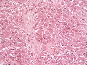
| Caption = Subependymal giant cell astrocytoma [[H&E stain]]. | |||
| Synonyms = SEGA | |||
| Micro = | |||
| Subtypes = | |||
| LMDDx = [[ganglioglioma]], [[pleomorphic xanthoastrocytoma]], [[glioblastoma]] | |||
| Stains = | |||
| IHC = GFAP +ve | |||
| EM = | |||
| Molecular = | |||
| IF = | |||
| Gross = | |||
| Grossing = | |||
| Site = brain - usu. wall of ventricles | |||
| Assdx = | |||
| Syndromes = | |||
| Clinicalhx = | |||
| Signs = | |||
| Symptoms = | |||
| Prevalence = rare - esp. in young adults | |||
| Bloodwork = | |||
| Rads = | |||
| Endoscopy = | |||
| Prognosis = good (WHO Grade I) | |||
| Other = | |||
| ClinDDx = | |||
| Tx = | |||
}} | |||
'''Subependymal giant cell astrocytoma''', abbreviated '''SEGA''', is a low-grade astrocytoma associated with [[tuberous sclerosis complex]]. | '''Subependymal giant cell astrocytoma''', abbreviated '''SEGA''', is a low-grade astrocytoma associated with [[tuberous sclerosis complex]]. | ||
==General== | ==General== | ||
*Associated with [[tuberous sclerosis complex]] (TSC).<ref name=pmid21455842>{{Cite journal | last1 = Grajkowska | first1 = W. | last2 = Kotulska | first2 = K. | last3 = Jurkiewicz | first3 = E. | last4 = Roszkowski | first4 = M. | last5 = Daszkiewicz | first5 = P. | last6 = Jóźwiak | first6 = S. | last7 = Matyja | first7 = E. | title = Subependymal giant cell astrocytomas with atypical histological features mimicking malignant gliomas. | journal = Folia Neuropathol | volume = 49 | issue = 1 | pages = 39-46 | month = | year = 2011 | doi = | PMID = 21455842 }}</ref> | *Associated with [[tuberous sclerosis complex]] (TSC).<ref name=pmid21455842>{{Cite journal | last1 = Grajkowska | first1 = W. | last2 = Kotulska | first2 = K. | last3 = Jurkiewicz | first3 = E. | last4 = Roszkowski | first4 = M. | last5 = Daszkiewicz | first5 = P. | last6 = Jóźwiak | first6 = S. | last7 = Matyja | first7 = E. | title = Subependymal giant cell astrocytomas with atypical histological features mimicking malignant gliomas. | journal = Folia Neuropathol | volume = 49 | issue = 1 | pages = 39-46 | month = | year = 2011 | doi = | PMID = 21455842 }}</ref> | ||
** 6-14% of all TSC patients will develop a SEGA. | |||
*Associated with epilepsy. | |||
*WHO Grade I. | *WHO Grade I. | ||
| Line 8: | Line 42: | ||
*Well-demarcated. | *Well-demarcated. | ||
*Often projecting into a ventricle. | *Often projecting into a ventricle. | ||
*May be calcified | |||
*Circumscribed tumour. | |||
<gallery> | <gallery> | ||
| Line 16: | Line 52: | ||
==Microscopic== | ==Microscopic== | ||
Features:<ref name=upmc_case179/><ref name=pmid9595853>{{Cite journal | last1 = Taraszewska | first1 = A. | last2 = Kroh | first2 = H. | last3 = Majchrowski | first3 = A. | title = Subependymal giant cell astrocytoma: clinical, histologic and immunohistochemical characteristic of 3 cases. | journal = Folia Neuropathol | volume = 35 | issue = 3 | pages = 181-6 | month = | year = 1997 | doi = | PMID = 9595853 }}</ref> | Features:<ref name=upmc_case179/><ref name=pmid9595853>{{Cite journal | last1 = Taraszewska | first1 = A. | last2 = Kroh | first2 = H. | last3 = Majchrowski | first3 = A. | title = Subependymal giant cell astrocytoma: clinical, histologic and immunohistochemical characteristic of 3 cases. | journal = Folia Neuropathol | volume = 35 | issue = 3 | pages = 181-6 | month = | year = 1997 | doi = | PMID = 9595853 }}</ref> | ||
*Giant cells with nuclear atypia ("bizarre cells"). | *Giant cells with nuclear atypia ("bizarre cells", "ganglioid cells"). | ||
**[[Vesicular nuclei]]. | **[[Vesicular nuclei]]. | ||
**[[Nuclear pseudoinclusions]].<ref name=upmc179>URL: [http://path.upmc.edu/cases/case179/micro.html http://path.upmc.edu/cases/case179/micro.html]. Accessed on: 8 January 2012.</ref> | **[[Nuclear pseudoinclusions]].<ref name=upmc179>URL: [http://path.upmc.edu/cases/case179/micro.html http://path.upmc.edu/cases/case179/micro.html]. Accessed on: 8 January 2012.</ref> | ||
*Glassy eosinophilic cytoplasm. | *Glassy eosinophilic cytoplasm. | ||
*Elongated cells in a fibrillary background. | |||
*Abundant [[mast cell]]s.<ref name=upmc179>URL: [http://path.upmc.edu/cases/case179/micro.html http://path.upmc.edu/cases/case179/micro.html]. Accessed on: 8 January 2012.</ref> | *Abundant [[mast cell]]s.<ref name=upmc179>URL: [http://path.upmc.edu/cases/case179/micro.html http://path.upmc.edu/cases/case179/micro.html]. Accessed on: 8 January 2012.</ref> | ||
*Lymphocytic infiltrates. | |||
*Endothelial proliferations and/or necrosis are not a sign of malignancy. | |||
===Images=== | ===Images=== | ||
| Line 38: | Line 77: | ||
*Vimentin +ve. (100%) | *Vimentin +ve. (100%) | ||
*S100 +ve. (100%) | *S100 +ve. (100%) | ||
* [[TTF-1]] (7 out of 7).<ref name=pmid25669749>{{Cite journal | last1 = Hewer | first1 = E. | last2 = Vajtai | first2 = I. | title = Consistent nuclear expression of thyroid transcription factor 1 in subependymal giant cell astrocytomas suggests lineage-restricted histogenesis. | journal = Clin Neuropathol | volume = 34 | issue = 3 | pages = 128-31 | month = | year = | doi = 10.5414/NP300818 | PMID = 25669749 }}</ref> | *Neurofilament +/-ve (ganglionic component). | ||
*Synaptophysin +/-ve (ganglionic component).. | |||
*[[TTF-1]] (7 out of 7).<ref name=pmid25669749>{{Cite journal | last1 = Hewer | first1 = E. | last2 = Vajtai | first2 = I. | title = Consistent nuclear expression of thyroid transcription factor 1 in subependymal giant cell astrocytomas suggests lineage-restricted histogenesis. | journal = Clin Neuropathol | volume = 34 | issue = 3 | pages = 128-31 | month = | year = | doi = 10.5414/NP300818 | PMID = 25669749 }}</ref> | |||
* MIB-1 usu. low (1-5%). | |||
==See also== | ==See also== | ||
| Line 48: | Line 90: | ||
[[Category:Diagnosis]] | [[Category:Diagnosis]] | ||
[[Category:Neuropathology tumours]] | [[Category:Neuropathology tumours]] | ||
[[Category:WHO grade I tumours]] | |||
Revision as of 14:55, 8 October 2015
| Subependymal giant cell astrocytoma | |
|---|---|
| Diagnosis in short | |
 Subependymal giant cell astrocytoma H&E stain. | |
|
| |
| Synonyms | SEGA |
| LM DDx | ganglioglioma, pleomorphic xanthoastrocytoma, glioblastoma |
| IHC | GFAP +ve |
| Site | brain - usu. wall of ventricles |
|
| |
| Prevalence | rare - esp. in young adults |
| Prognosis | good (WHO Grade I) |
Subependymal giant cell astrocytoma, abbreviated SEGA, is a low-grade astrocytoma associated with tuberous sclerosis complex.
General
- Associated with tuberous sclerosis complex (TSC).[1]
- 6-14% of all TSC patients will develop a SEGA.
- Associated with epilepsy.
- WHO Grade I.
Gross/radiology
- Well-demarcated.
- Often projecting into a ventricle.
- May be calcified
- Circumscribed tumour.
Microscopic
- Giant cells with nuclear atypia ("bizarre cells", "ganglioid cells").
- Glassy eosinophilic cytoplasm.
- Elongated cells in a fibrillary background.
- Abundant mast cells.[4]
- Lymphocytic infiltrates.
- Endothelial proliferations and/or necrosis are not a sign of malignancy.
Images
www:
IHC
- GFAP +ve. (50%)
- Vimentin +ve. (100%)
- S100 +ve. (100%)
- Neurofilament +/-ve (ganglionic component).
- Synaptophysin +/-ve (ganglionic component)..
- TTF-1 (7 out of 7).[6]
- MIB-1 usu. low (1-5%).
See also
References
- ↑ Grajkowska, W.; Kotulska, K.; Jurkiewicz, E.; Roszkowski, M.; Daszkiewicz, P.; Jóźwiak, S.; Matyja, E. (2011). "Subependymal giant cell astrocytomas with atypical histological features mimicking malignant gliomas.". Folia Neuropathol 49 (1): 39-46. PMID 21455842.
- ↑ 2.0 2.1 URL: http://path.upmc.edu/cases/case179.html. Accessed on: 29 July 2011.
- ↑ 3.0 3.1 Taraszewska, A.; Kroh, H.; Majchrowski, A. (1997). "Subependymal giant cell astrocytoma: clinical, histologic and immunohistochemical characteristic of 3 cases.". Folia Neuropathol 35 (3): 181-6. PMID 9595853.
- ↑ 4.0 4.1 URL: http://path.upmc.edu/cases/case179/micro.html. Accessed on: 8 January 2012.
- ↑ Hirose, T.; Scheithauer, BW.; Lopes, MB.; Gerber, HA.; Altermatt, HJ.; Hukee, MJ.; VandenBerg, SR.; Charlesworth, JC. (1995). "Tuber and subependymal giant cell astrocytoma associated with tuberous sclerosis: an immunohistochemical, ultrastructural, and immunoelectron and microscopic study.". Acta Neuropathol 90 (4): 387-99. PMID 8546029.
- ↑ Hewer, E.; Vajtai, I.. "Consistent nuclear expression of thyroid transcription factor 1 in subependymal giant cell astrocytomas suggests lineage-restricted histogenesis.". Clin Neuropathol 34 (3): 128-31. doi:10.5414/NP300818. PMID 25669749.


