Difference between revisions of "Lymphangioleiomyomatosis"
Jump to navigation
Jump to search
(+infobox) |
|||
| Line 1: | Line 1: | ||
{{ Infobox diagnosis | {{ Infobox diagnosis | ||
| Name = {{PAGENAME}} | | Name = {{PAGENAME}} | ||
| Image = | | Image = Lymphangioleiomyomatosis - intermed mag.jpg | ||
| Width = | | Width = | ||
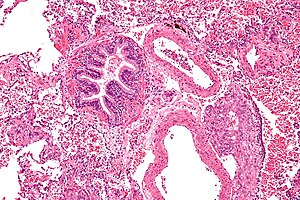
| Caption = | | Caption = Lymphangioleiomyomatosis. [[H&E stain]]. | ||
| Synonyms = lymphangiomyomatosis | | Synonyms = lymphangiomyomatosis | ||
| Micro = spindle cells with small nuclei + larger epithelioid cells with clear cytoplasm and round nuclei, cyst formation, thick arterial walls | | Micro = spindle cells with small nuclei + larger epithelioid cells with clear cytoplasm and round nuclei, cyst formation, thick arterial walls | ||
Revision as of 06:03, 9 December 2014
| Lymphangioleiomyomatosis | |
|---|---|
| Diagnosis in short | |
 Lymphangioleiomyomatosis. H&E stain. | |
|
| |
| Synonyms | lymphangiomyomatosis |
|
| |
| LM | spindle cells with small nuclei + larger epithelioid cells with clear cytoplasm and round nuclei, cyst formation, thick arterial walls |
| IHC | HMB-45 +ve, ER +ve, PR +ve, SMA +ve |
| Site | lung - see medical lung diseases |
|
| |
| Syndromes | tuberous sclerosis |
|
| |
| Clinical history | almost always women of childbearing age, recurrent pneumothorax |
| Symptoms | dyspnea |
| Prevalence | very rare |
| Radiology | bullae/thin walled cysts distributed in all lung fields, lymphadenopathy |
| Clin. DDx | eosinophilic granuloma, usual interstitial pneumonia, emphysema |
| Treatment | lung transplantation |
Lymphangioleiomyomatosis, abbreviated LAM, is a rare lung pathology that predominantly afflicits women of childbearing age.
It is also known as lymphangiomyomatosis.
General
- Clinical: dyspnea, recurrent pneumothorax.
- May be an indication for lung transplantation.
- Non-neoplastic muscle proliferation versus tumour that can metastasize.[1]
Notes:
- Considered to be a PEComa.
Epidemiology
- Associated with angiomyolipomas.[2]
- Associated with tuberous sclerosis[2] - abnormality in same gene (TSC2).
- Usually affects women - primarily in childbearing years; case reports of LAM in men - usu. with TSC.[3]
- Rare.
Radiology
- Bullae/thin walled cysts - distributed in all lung fields.
- Lymphadenopathy.
Radiologic DDx (of cysts):
- Eosinophilic granuloma - associated with smoking.
- Usual interstitial pneumonia (UIP).
- Emphysema.
Microscopic
Features:[4]
- Spindle cells with small nuclei + larger epithelioid cells with clear cytoplasm and round nuclei.
- Cyst formation.
- Thick arterial walls.
Images
www:
IHC
- HMB-45 +ve.
- ER +ve.
- PR +ve.
- SMA +ve.
See also
References
- ↑ Taveira-DaSilva, AM.; Pacheco-Rodriguez, G.; Moss, J. (Mar 2010). "The natural history of lymphangioleiomyomatosis: markers of severity, rate of progression and prognosis.". Lymphat Res Biol 8 (1): 9-19. doi:10.1089/lrb.2009.0024. PMID 20235883.
- ↑ 2.0 2.1 http://emedicine.medscape.com/article/299545-overview
- ↑ Schiavina, M.; Di Scioscio, V.; Contini, P.; Cavazza, A.; Fabiani, A.; Barberis, M.; Bini, A.; Altimari, A. et al. (Jul 2007). "Pulmonary lymphangioleiomyomatosis in a karyotypically normal man without tuberous sclerosis complex.". Am J Respir Crit Care Med 176 (1): 96-8. doi:10.1164/rccm.200610-1408CR. PMID 17431222.
- ↑ http://emedicine.medscape.com/article/299545-diagnosis


