Difference between revisions of "Steatocystoma"
Jump to navigation
Jump to search
(+cat.) |
(split out) |
||
| Line 1: | Line 1: | ||
# | '''Steatocystoma''' is a rare benign [[dermal cyst]]. | ||
==General== | |||
*Benign. | |||
*Typically adults. | |||
*Usually on the trunk. | |||
*May be genetic; known as ''steatocystoma multiplex''.<ref name=omim184500>{{OMIM|184500}}</ref> | |||
**Classically autosomal dominant.<ref>URL: [http://path.upmc.edu/cases/case674/dx.html http://path.upmc.edu/cases/case674/dx.html]. Accessed on: 29 January 2012.</ref> | |||
==Microscopic== | |||
Features:<ref name=Ref_Derm312>{{Ref Derm|312}}</ref> | |||
*Cyst lined by squamous epithelium with: | |||
*#Corrugated eosinophilic lining - '''key feature'''. | |||
*#*Similar appearance to compact keratin (hyperkeratosis). | |||
*#*Described as a ''hyaline cuticle''.<ref>URL: [http://path.upmc.edu/cases/case674/dx.html http://path.upmc.edu/cases/case674/dx.html]. Accessed on: 29 January 2012.</ref> | |||
*#'''No''' granular cell layer. | |||
===Images=== | |||
<gallery> | |||
Image:SkinTumors-P6260388.JPG | Steatocystoma. (WC) | |||
Image:Steatocystoma_-_low_mag.jpg | Steatocystoma - low mag. (WC/Nephron) | |||
Image:Steatocystoma_-_intermed_mag.jpg | Steatocystoma - intermed. mag. (WC/Nephron) | |||
</gallery> | |||
www: | |||
*[http://www.flickr.com/photos/santoshpath/4788590105/in/photostream/ Steatocystoma - low mag. (flickr.com)]. | |||
**[http://www.flickr.com/photos/santoshpath/4788590109/ Steatocystoma - high mag. (flickr.com)]. | |||
*[http://path.upmc.edu/cases/case674/images/fig03.jpg Steatocystoma (upmc.edu)].<ref>URL: [http://path.upmc.edu/cases/case674.html http://path.upmc.edu/cases/case674.html]. Accessed on: 29 January 2012.</ref> | |||
==See also== | |||
*[[Dermal cysts]]. | |||
==References== | |||
{{Reflist|1}} | |||
[[Category:Diagnosis]] | [[Category:Diagnosis]] | ||
[[Category:Dermal cysts]] | |||
Revision as of 22:46, 16 February 2014
Steatocystoma is a rare benign dermal cyst.
General
- Benign.
- Typically adults.
- Usually on the trunk.
- May be genetic; known as steatocystoma multiplex.[1]
- Classically autosomal dominant.[2]
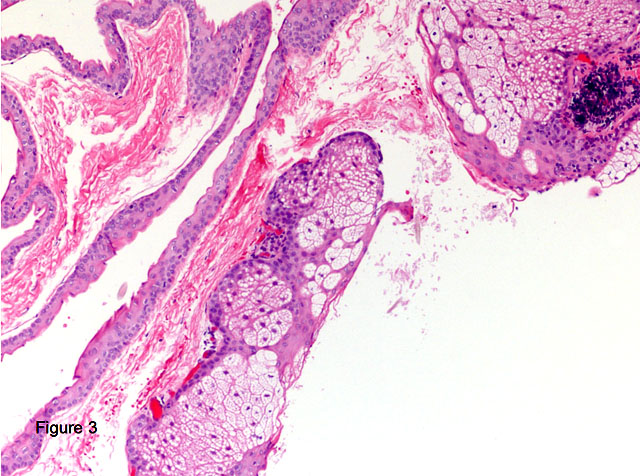
Microscopic
Features:[3]
- Cyst lined by squamous epithelium with:
- Corrugated eosinophilic lining - key feature.
- Similar appearance to compact keratin (hyperkeratosis).
- Described as a hyaline cuticle.[4]
- No granular cell layer.
- Corrugated eosinophilic lining - key feature.
Images
- SkinTumors-P6260388.JPG
Steatocystoma. (WC)
- Steatocystoma - low mag.jpg
Steatocystoma - low mag. (WC/Nephron)
- Steatocystoma - intermed mag.jpg
Steatocystoma - intermed. mag. (WC/Nephron)
www:
See also
References
- ↑ Online 'Mendelian Inheritance in Man' (OMIM) 184500
- ↑ URL: http://path.upmc.edu/cases/case674/dx.html. Accessed on: 29 January 2012.
- ↑ Busam, Klaus J. (2009). Dermatopathology: A Volume in the Foundations in Diagnostic Pathology Series (1st ed.). Saunders. pp. 312. ISBN 978-0443066542.
- ↑ URL: http://path.upmc.edu/cases/case674/dx.html. Accessed on: 29 January 2012.
- ↑ URL: http://path.upmc.edu/cases/case674.html. Accessed on: 29 January 2012.