Difference between revisions of "Myxomatous degeneration"
Jump to navigation
Jump to search
(+cat.) |
(split out) |
||
| Line 1: | Line 1: | ||
'''Myxomatous degeneration''' of [[heart valves]] is rare benign condition that is typically seen in the mitral valve, and may be associated with various genetic conditions. | |||
==General== | |||
*Usually affects the mitral valve. | |||
*Female > male,<ref>URL: [http://emedicine.medscape.com/article/759004-overview http://emedicine.medscape.com/article/759004-overview]. Accessed on: 8 June 2010.</ref> disputed by Toronto data.<ref name=leong>{{cite journal |author=Leong SW, Soor GS, Butany J, Henry J, Thangaroopan M, Leask RL |title=Morphological findings in 192 surgically excised native mitral valves |journal=Can J Cardiol |volume=22 |issue=12 |pages=1055-61 |year=2006 |month=October |pmid=17036100 |doi= |url=}}</ref> | |||
*Associated with [[Marfan's syndrome]] and [[Turner syndrome]] (Monosomy X).<ref name=pmid779595>{{cite journal |author=Wigle ED, Rakowski H, Ranganathan N, Silver MC |title=Mitral valve prolapse |journal=Annu. Rev. Med. |volume=27 |issue= |pages=165–80 |year=1976 |pmid=779595 |doi=10.1146/annurev.me.27.020176.001121 |url=}}</ref> | |||
==Gross== | |||
Features:<ref name=Ref_PBoD591>{{Ref PBoD|591}}</ref> | |||
*No commissural fusion. | |||
**Commissural fusion typical of rheumatic heart disease. | |||
*Thickened. | |||
*Rubbery consistency. | |||
*Reactive/secondary changes. | |||
**Fibrosis due to prolapse/abnormal contact of valve with other structures. | |||
**Clots/organized thrombus - due to stasis. | |||
==Microscopic== | |||
*Thinning of ''fibrosa layer''. | |||
*Thickening of ''spongiosa layer'' with mucoid (myxomatous) material. (key feature). | |||
*+/-Secondary changes (due to valvular dysfunction): thrombi, fibrosis. | |||
==Staining== | |||
*Movat stain. | |||
**Acid fuchsin, alcian blue, crocein scarlet, elastic hematoxylin, pathology consultation, and saffron.<ref>URL: [http://www.mayomedicallaboratories.com/test-catalog/Overview/9832 http://www.mayomedicallaboratories.com/test-catalog/Overview/9832]. Accessed on: 8 June 2010.</ref><ref name=penn_med>Modified Movat's Pentachrome Stain. University Penn Medicine. URL: [http://www.med.upenn.edu/mcrc/histology_core/movat.shtml http://www.med.upenn.edu/mcrc/histology_core/movat.shtml]. Accessed on: January 29, 2009.</ref> | |||
Interpretation of Movat stain:<ref name=penn_med/> | |||
*Black = nuclei and elastic fibers. | |||
*Yellow = collagen and reticular fibers. | |||
*Blue = mucin, ground substance. | |||
*Red (intense) = fibrin. | |||
*Red = muscle. | |||
===Image=== | |||
<gallery> | |||
Image:Myxomatous_aortic_valve.jpg | Myxomatous valve. [[Movat stain]]. (WC/Nephron) | |||
</gallery> | |||
==See also== | |||
*[[Heart valves]]. | |||
==References== | |||
{{reflist|2}} | |||
[[Category:Diagnosis]] | [[Category:Diagnosis]] | ||
[[Category:Heart valves]] | |||
Revision as of 05:50, 5 April 2015
Myxomatous degeneration of heart valves is rare benign condition that is typically seen in the mitral valve, and may be associated with various genetic conditions.
General
- Usually affects the mitral valve.
- Female > male,[1] disputed by Toronto data.[2]
- Associated with Marfan's syndrome and Turner syndrome (Monosomy X).[3]
Gross
Features:[4]
- No commissural fusion.
- Commissural fusion typical of rheumatic heart disease.
- Thickened.
- Rubbery consistency.
- Reactive/secondary changes.
- Fibrosis due to prolapse/abnormal contact of valve with other structures.
- Clots/organized thrombus - due to stasis.
Microscopic
- Thinning of fibrosa layer.
- Thickening of spongiosa layer with mucoid (myxomatous) material. (key feature).
- +/-Secondary changes (due to valvular dysfunction): thrombi, fibrosis.
Staining
- Movat stain.
Interpretation of Movat stain:[6]
- Black = nuclei and elastic fibers.
- Yellow = collagen and reticular fibers.
- Blue = mucin, ground substance.
- Red (intense) = fibrin.
- Red = muscle.
Image
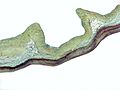
Myxomatous valve. Movat stain. (WC/Nephron)

See also
References
- ↑ URL: http://emedicine.medscape.com/article/759004-overview. Accessed on: 8 June 2010.
- ↑ Leong SW, Soor GS, Butany J, Henry J, Thangaroopan M, Leask RL (October 2006). "Morphological findings in 192 surgically excised native mitral valves". Can J Cardiol 22 (12): 1055-61. PMID 17036100.
- ↑ Wigle ED, Rakowski H, Ranganathan N, Silver MC (1976). "Mitral valve prolapse". Annu. Rev. Med. 27: 165–80. doi:10.1146/annurev.me.27.020176.001121. PMID 779595.
- ↑ Cotran, Ramzi S.; Kumar, Vinay; Fausto, Nelson; Nelso Fausto; Robbins, Stanley L.; Abbas, Abul K. (2005). Robbins and Cotran pathologic basis of disease (7th ed.). St. Louis, Mo: Elsevier Saunders. pp. 591. ISBN 0-7216-0187-1.
- ↑ URL: http://www.mayomedicallaboratories.com/test-catalog/Overview/9832. Accessed on: 8 June 2010.
- ↑ 6.0 6.1 Modified Movat's Pentachrome Stain. University Penn Medicine. URL: http://www.med.upenn.edu/mcrc/histology_core/movat.shtml. Accessed on: January 29, 2009.