Difference between revisions of "Giant cell arteritis"
Jump to navigation
Jump to search
| Line 55: | Line 55: | ||
*Destruction of arterial wall, e.g. fibrinoid necrosis (pink anucleate arterial wall). | *Destruction of arterial wall, e.g. fibrinoid necrosis (pink anucleate arterial wall). | ||
Image( | ===Images=== | ||
<gallery> | |||
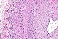
Image: Giant cell arteritis -- very low mag.jpg | GCA - very low mag. (WC) | |||
Image: Giant cell arteritis -- low mag.jpg | GCA - low mag. (WC) | |||
Image: Giant cell arteritis -- intermed mag.jpg | GCA - intermed. mag. (WC) | |||
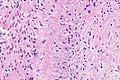
Image: Giant cell arteritis -- high mag.jpg | GCA - high mag. (WC) | |||
Image: Giant cell arteritis - alt -- intermed mag.jpg | GCA - intermed mag. (WC) | |||
Image: Giant cell arteritis - alt -- high mag.jpg | GCA - high mag. (WC) | |||
</gallery> | |||
www: | |||
*[http://www.djo.harvard.edu/files/5077_728.jpg GCA (harvard.edu)]. | *[http://www.djo.harvard.edu/files/5077_728.jpg GCA (harvard.edu)]. | ||
*[http://path.upmc.edu/cases/case646.html GCA - several images (upmc.edu)]. | *[http://path.upmc.edu/cases/case646.html GCA - several images (upmc.edu)]. | ||
Revision as of 08:01, 17 December 2014
| Giant cell arteritis | |
|---|---|
| Diagnosis in short | |
 Giant cell arteritis. H&E stain. | |
|
| |
| Synonyms | temporal arteritis |
|
| |
| LM | large artery with intramural inflammatory cells (often granulomatous); destruction of arterial wall, i.e. fibrinoid necrosis (pink anucleate arterial wall) |
| Site | large blood vessels - see vasculitides |
|
| |
| Clinical history | patient older than 50 years |
| Signs | loss of vision, weight loss, chills, fever |
| Symptoms | headache, double vision, scalp tenderness |
| Prevalence | uncommon |
| Blood work | ESR elevated |
| Prognosis | good if treated |
| Clin. DDx | other causes of headache |
| Treatment | steroids |
Giant cell arteritis (abbreviated GCA), also known as temporal arteritis, is a type of large vessel vasculitis.
General
- Classically afflicts the temporal artery.
Clinical features:
- Classic finding: jaw claudication, in a patient older than 50 years.
- Other findings: headache, vision loss or diplopia, scalp tenderness, polymyalgia, weight loss, chills, fever.
Work-up:
- CRP, ESR, temporal artery biopsy.
- ESR normal (>50 years old): <20 mm/hr males, <30 mm/hr females.[1]
Treatment:
- Treat right away with high dose steroids.
- Biopsy is confirmatory.
Microscopic
Features:
- Artery with intramural inflammatory cells.
- Classically granulomatous inflammation.
- Granulomas not required for the diagnosis!
- Classically granulomatous inflammation.
- Destruction of arterial wall, e.g. fibrinoid necrosis (pink anucleate arterial wall).
Images
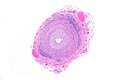
GCA - very low mag. (WC)
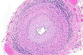
GCA - low mag. (WC)
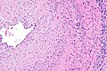
GCA - intermed. mag. (WC)
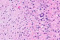
GCA - high mag. (WC)
GCA - intermed mag. (WC)
GCA - high mag. (WC)


www:
Sign out
Negative
TEMPORAL ARTERY, LEFT, BIOPSY:
- MEDIUM SIZE ARTERY WITHOUT PATHOLOGIC DIAGNOSIS, SEE COMMENT.

COMMENT:
A negative biopsy does not rule out the possibility of giant cell (temporal)
arteritis, as this may be a focal disorder. The clinical management is
dependent upon the clinical impression.
See also
References
- ↑ URL: http://www.nlm.nih.gov/medlineplus/ency/article/003638.htm. Accessed on: 17 August 2012.